8C30

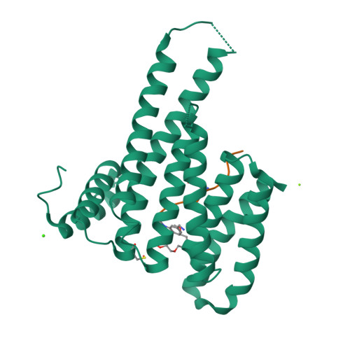
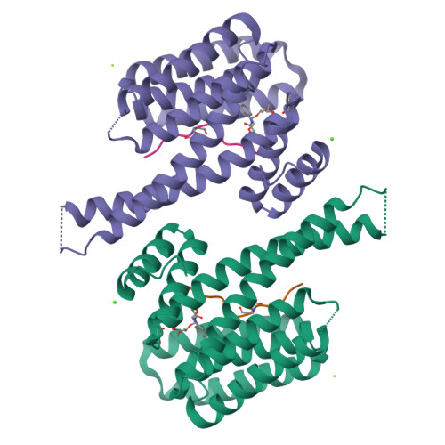
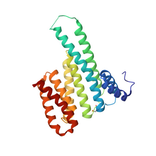
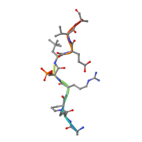
Crystal structure of 14-3-3 in complex with PyrinpS242 and a protein/peptide interface fragment


Experimental Data Snapshot

  • Method: X-RAY DIFFRACTION
  • Resolution: 1.40 Å
  • R-Value Free: 0.204 
  • R-Value Work: 0.179 

Starting Model: experimental
View more details

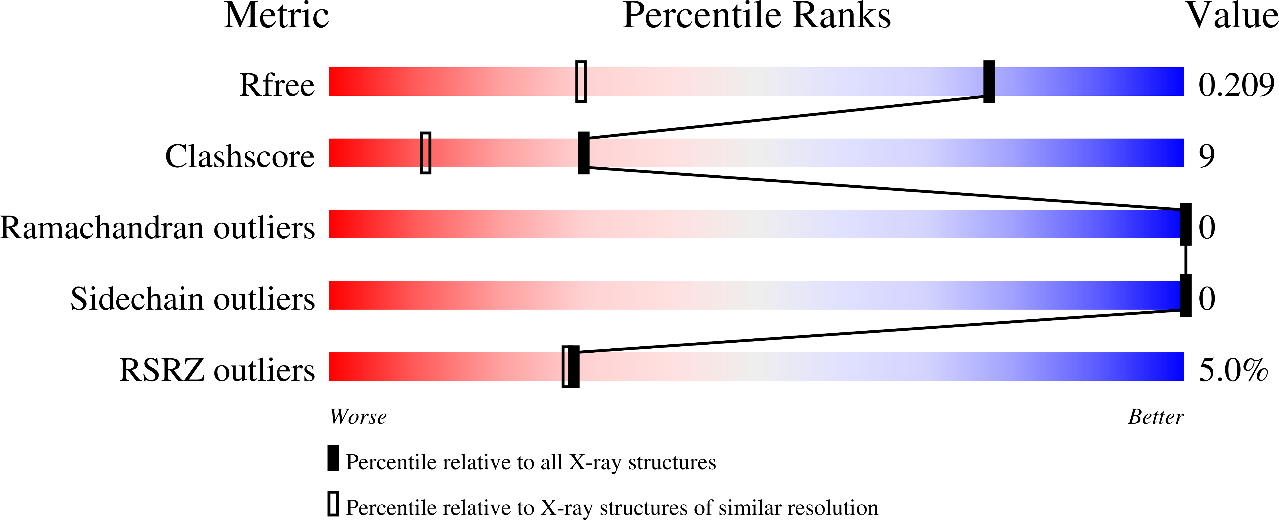
wwPDB Validation   3D Report Full Report


Ligand Structure Quality Assessment 


This is version 1.2 of the entry. See complete history


Literature

Crystal structure and ligandability of the 14-3-3/pyrin interface.

Lau, R.Hann, M.M.Ottmann, C.

(2023) Biochem Biophys Res Commun 651: 1-7

  • DOI: https://doi.org/10.1016/j.bbrc.2023.02.013
  • Primary Citation of Related Structures:  
    8C28, 8C2D, 8C2Y, 8C30

  • PubMed Abstract: 

    Overactivation of Pyrin is the cause of the inflammatory diseases Mediterranean Fever and Pyrin-associated autoinflammation with neutrophilic dermatosis (PAAND). Binding of 14-3-3 proteins reduces the pro-inflammatory activity of Pyrin, hence small molecules that stabilize the Pyrin/14-3-3 complex could convey an anti-inflammatory effect. We have solved the atomic resolution crystal structures of phosphorylated peptides derived from PyrinpS208 and PyrinpS242 - the two principle 14-3-3 binding sites in Pyrin - in complex with 14-3-3 and analyzed the ligandability of these protein-peptide interfaces by crystal-based fragment soaking. The complex between 14-3-3 and PyrinpS242 appears to be much more amenable for small-molecule binding than that of 14-3-3/PyrinpS208. Consequently, only for the 14-3-3/PyrinpS242 complex could we find an interface-binding fragment, validating protein crystallography and fragment soaking as a method to evaluate the ligandability of protein surfaces.


  • Organizational Affiliation

    Laboratory of Chemical Biology, Department of Biomedical Engineering and Institute for Complex Molecular Systems, Technische Universiteit Eindhoven, Den Dolech 2, 5612 AZ, Eindhoven, the Netherlands.


Macromolecules
Find similar proteins by:  (by identity cutoff)  |  3D Structure
Entity ID: 1
MoleculeChains Sequence LengthOrganismDetailsImage
14-3-3 protein sigmaA [auth AAA]236Homo sapiensMutation(s): 0 
Gene Names: SFNHME1
UniProt & NIH Common Fund Data Resources
Find proteins for P31947 (Homo sapiens)
Explore P31947 
Go to UniProtKB:  P31947
PHAROS:  P31947
GTEx:  ENSG00000175793 
Entity Groups  
Sequence Clusters30% Identity50% Identity70% Identity90% Identity95% Identity100% Identity
UniProt GroupP31947
Sequence Annotations
Expand
  • Reference Sequence

Find similar proteins by:  Sequence   |   3D Structure  

Entity ID: 2
MoleculeChains Sequence LengthOrganismDetailsImage
PyrinB [auth PPP]12Homo sapiensMutation(s): 0 
UniProt & NIH Common Fund Data Resources
Find proteins for O15553 (Homo sapiens)
Explore O15553 
Go to UniProtKB:  O15553
PHAROS:  O15553
GTEx:  ENSG00000103313 
Entity Groups  
Sequence Clusters30% Identity50% Identity70% Identity90% Identity95% Identity100% Identity
UniProt GroupO15553
Sequence Annotations
Expand
  • Reference Sequence
Small Molecules
Modified Residues  2 Unique
IDChains TypeFormula2D DiagramParent
CSO
Query on CSO
A [auth AAA]L-PEPTIDE LINKINGC3 H7 N O3 SCYS
SEP
Query on SEP
B [auth PPP]L-PEPTIDE LINKINGC3 H8 N O6 PSER
Experimental Data & Validation

Experimental Data

  • Method: X-RAY DIFFRACTION
  • Resolution: 1.40 Å
  • R-Value Free: 0.204 
  • R-Value Work: 0.179 
  • Space Group: C 2 2 21
Unit Cell:
Length ( Å )Angle ( ˚ )
a = 82.142α = 90
b = 111.969β = 90
c = 62.477γ = 90
Software Package:
Software NamePurpose
REFMACrefinement
DIALSdata reduction
DIALSdata scaling
PHASERphasing

Structure Validation

View Full Validation Report


Ligand Structure Quality Assessment 


Entry History & Funding Information

Deposition Data


Funding OrganizationLocationGrant Number
Not funded--

Revision History  (Full details and data files)

  • Version 1.0: 2024-01-10
    Type: Initial release
  • Version 1.1: 2024-11-06
    Changes: Structure summary
  • Version 1.2: 2025-01-22
    Changes: Database references