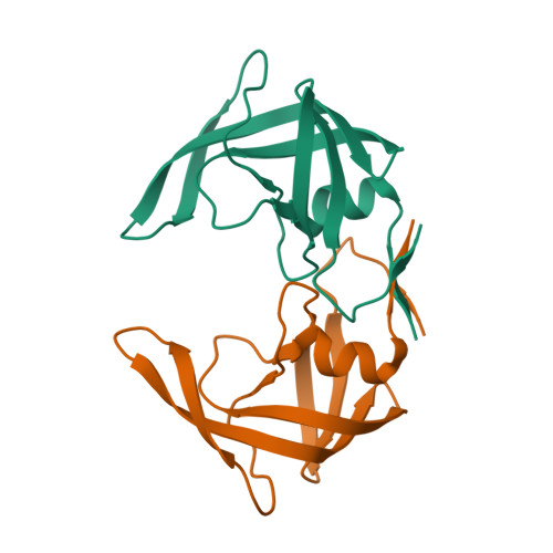
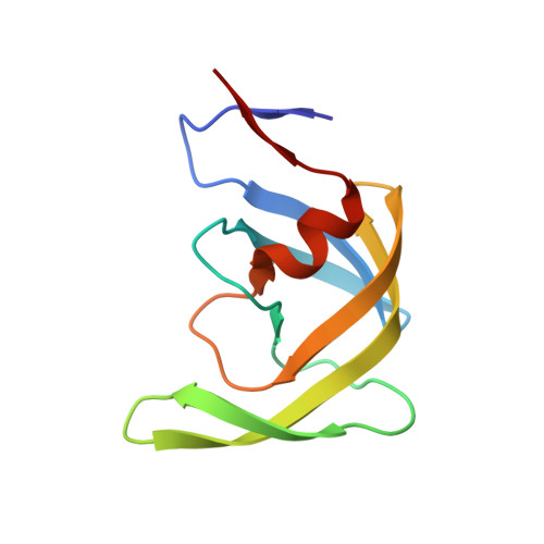
8DCI

Crystal Structure of a highly resistant HIV-1 protease Clinical isolate PR10x (inhibitor-free)


Experimental Data Snapshot

  • Method: X-RAY DIFFRACTION
  • Resolution: 1.62 Å
  • R-Value Free: 0.227 
  • R-Value Work: 0.189 
  • R-Value Observed: 0.190 

Starting Model: experimental
View more details

wwPDB Validation   3D Report Full Report


This is version 1.1 of the entry. See complete history


Literature

HIV-1 protease with 10 lopinavir and darunavir resistance mutations exhibits altered inhibition, structural rearrangements and extreme dynamics.

Wong-Sam, A.Wang, Y.F.Kneller, D.W.Kovalevsky, A.Y.Ghosh, A.K.Harrison, R.W.Weber, I.T.

(2022) J Mol Graph Model 117: 108315-108315

  • DOI: https://doi.org/10.1016/j.jmgm.2022.108315
  • Primary Citation of Related Structures:  
    8DCH, 8DCI

  • PubMed Abstract: 

    Antiretroviral drug resistance is a therapeutic obstacle for people with HIV. HIV protease inhibitors darunavir and lopinavir are recommended for resistant infections. We characterized a protease mutant (PR10x) derived from a highly resistant clinical isolate including 10 mutations associated with resistance to lopinavir and darunavir. Compared to the wild-type protease, PR10x exhibits ∼3-fold decrease in catalytic efficiency and K i values of 2-3 orders of magnitude worse for darunavir, lopinavir, and potent investigational inhibitor GRL-519. Crystal structures of the mutant were solved in a ligand-free form and in complex with GRL-519. The structures show altered interactions in the active site, flap-core interface, hydrophobic core, hinge region, and 80s loop compared to the corresponding wild-type protease structures. The ligand-free crystal structure exhibits a highly curled flap conformation which may amplify drug resistance. Molecular dynamics simulations performed for 1 μs on ligand-free dimers showed extremely large fluctuations in the flaps for PR10x compared to equivalent simulations on PR with a single L76V mutation or wild-type protease. This analysis offers insight about the synergistic effects of mutations in highly resistant variants.


  • Organizational Affiliation

    Department of Biology, Georgia State University, Atlanta, GA, 30303, USA.


Macromolecules
Find similar proteins by:  (by identity cutoff)  |  3D Structure
Entity ID: 1
MoleculeChains Sequence LengthOrganismDetailsImage
Protease
A, B
99Human immunodeficiency virus 1Mutation(s): 23 
Gene Names: gag-pol
EC: 3.4.23.16
UniProt
Find proteins for P03367 (Human immunodeficiency virus type 1 group M subtype B (isolate BRU/LAI))
Explore P03367 
Go to UniProtKB:  P03367
Entity Groups  
Sequence Clusters30% Identity50% Identity70% Identity90% Identity95% Identity100% Identity
UniProt GroupP03367
Sequence Annotations
Expand
  • Reference Sequence
Experimental Data & Validation

Experimental Data

  • Method: X-RAY DIFFRACTION
  • Resolution: 1.62 Å
  • R-Value Free: 0.227 
  • R-Value Work: 0.189 
  • R-Value Observed: 0.190 
  • Space Group: P 21 21 21
Unit Cell:
Length ( Å )Angle ( ˚ )
a = 28.366α = 90
b = 81.351β = 90
c = 85.615γ = 90
Software Package:
Software NamePurpose
REFMACrefinement
HKL-2000data reduction
HKL-2000data scaling
PHASERphasing
PDB_EXTRACTdata extraction

Structure Validation

View Full Validation Report


Entry History & Funding Information

Deposition Data


Funding OrganizationLocationGrant Number
National Institutes of Health/National Cancer Institute (NIH/NCI)United StatesAI150461
National Institutes of Health/National Cancer Institute (NIH/NCI)United StatesAI150466
National Institutes of Health/National Cancer Institute (NIH/NCI)United Statesan NIH diversity supplement
Department of Energy (DOE, United States)United StatesW-31-109-Eng-38

Revision History  (Full details and data files)

  • Version 1.0: 2022-10-05
    Type: Initial release
  • Version 1.1: 2023-10-18
    Changes: Data collection, Refinement description