8DRJ

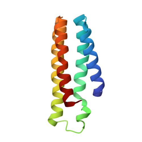
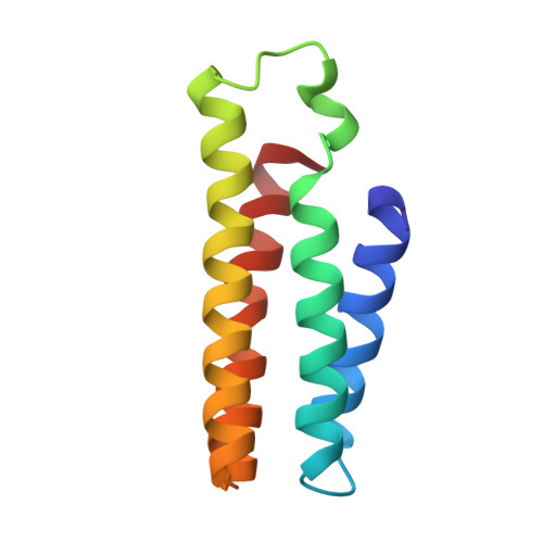
Apo B2 dimer (H60/H100/H104) formed in the presence of Cu(II)


Experimental Data Snapshot

  • Method: X-RAY DIFFRACTION
  • Resolution: 2.40 Å
  • R-Value Free: 0.283 
  • R-Value Work: 0.191 
  • R-Value Observed: 0.201 

Starting Model: experimental
View more details

wwPDB Validation   3D Report Full Report


Ligand Structure Quality Assessment 


This is version 1.2 of the entry. See complete history


Literature

Design of a Flexible, Zn-Selective Protein Scaffold that Displays Anti-Irving-Williams Behavior.

Choi, T.S.Tezcan, F.A.

(2022) J Am Chem Soc 144: 18090-18100

  • DOI: https://doi.org/10.1021/jacs.2c08050
  • Primary Citation of Related Structures:  
    8DRD, 8DRF, 8DRJ, 8DRL, 8DRM

  • PubMed Abstract: 

    Selective metal binding is a key requirement not only for the functions of natural metalloproteins but also for the potential applications of artificial metalloproteins in heterogeneous environments such as cells and environmental samples. The selection of transition-metal ions through protein design can, in principle, be achieved through the appropriate choice and the precise positioning of amino acids that comprise the primary metal coordination sphere. However, this task is made difficult by the intrinsic flexibility of proteins and the fact that protein design approaches generally lack the sub-Å precision required for the steric selection of metal ions. We recently introduced a flexible/probabilistic protein design strategy (MASCoT) that allows metal ions to search for optimal coordination geometry within a flexible, yet covalently constrained dimer interface. In an earlier proof-of-principle study, we used MASCoT to generate an artificial metalloprotein dimer, (AB) 2 , which selectively bound Co II and Ni II over Cu II (as well as other first-row transition-metal ions) through the imposition of a rigid octahedral coordination geometry, thus countering the Irving-Williams trend. In this study, we set out to redesign (AB) 2 to examine the applicability of MASCoT to the selective binding of other metal ions. We report here the design and characterization of a new flexible protein dimer, B 2 , which displays Zn II selectivity over all other tested metal ions including Cu II both in vitro and in cellulo . Selective, anti-Irving-Williams Zn II binding by B 2 is achieved through the formation of a unique trinuclear Zn coordination motif in which His and Glu residues are rigidly placed in a tetrahedral geometry. These results highlight the utility of protein flexibility in the design and discovery of selective binding motifs.


  • Organizational Affiliation

    Department of Chemistry and Biochemistry, University of California, San Diego, 9500 Gilman Drive, La Jolla, California 92093 United States.


Macromolecules
Find similar proteins by:  (by identity cutoff)  |  3D Structure
Entity ID: 1
MoleculeChains Sequence LengthOrganismDetailsImage
Soluble cytochrome b562A [auth C]106Escherichia coliMutation(s): 3 
Gene Names: cybC
UniProt
Find proteins for P0ABE7 (Escherichia coli)
Explore P0ABE7 
Go to UniProtKB:  P0ABE7
Entity Groups  
Sequence Clusters30% Identity50% Identity70% Identity90% Identity95% Identity100% Identity
UniProt GroupP0ABE7
Sequence Annotations
Expand
  • Reference Sequence
Find similar proteins by:  (by identity cutoff)  |  3D Structure
Entity ID: 2
MoleculeChains Sequence LengthOrganismDetailsImage
Soluble cytochrome b562B [auth A]106Escherichia coliMutation(s): 3 
Gene Names: cybC
UniProt
Find proteins for P0ABE7 (Escherichia coli)
Explore P0ABE7 
Go to UniProtKB:  P0ABE7
Entity Groups  
Sequence Clusters30% Identity50% Identity70% Identity90% Identity95% Identity100% Identity
UniProt GroupP0ABE7
Sequence Annotations
Expand
  • Reference Sequence
Experimental Data & Validation

Experimental Data

  • Method: X-RAY DIFFRACTION
  • Resolution: 2.40 Å
  • R-Value Free: 0.283 
  • R-Value Work: 0.191 
  • R-Value Observed: 0.201 
  • Space Group: C 1 2 1
Unit Cell:
Length ( Å )Angle ( ˚ )
a = 89.32α = 90
b = 48.02β = 96.18
c = 45.12γ = 90
Software Package:
Software NamePurpose
PHENIXrefinement
iMOSFLMdata reduction
Aimlessdata scaling
PHASERphasing

Structure Validation

View Full Validation Report


Ligand Structure Quality Assessment 


Entry History & Funding Information

Deposition Data


Funding OrganizationLocationGrant Number
National Institutes of Health/National Institute of General Medical Sciences (NIH/NIGMS)United StatesR01-GM138884

Revision History  (Full details and data files)

  • Version 1.0: 2022-10-12
    Type: Initial release
  • Version 1.1: 2023-10-18
    Changes: Data collection, Refinement description
  • Version 1.2: 2024-12-25
    Changes: Advisory, Derived calculations, Structure summary