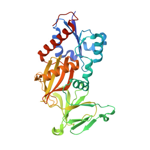
8G9E | pdb_00008g9e

Crystal structure of the catalytic domain of human diphosphoinositol pentakisphosphate kinase 2 (PPIP5K2) in complex with AMP-PNP and 5-(PCF2P)-IP5, an analog of 5-IP7

  • Classification: TRANSFERASE
  • Organism(s): Homo sapiens
  • Expression System: Escherichia coli
  • Mutation(s): No 

  • Deposited: 2023-02-21 Released: 2024-01-03 
  • Deposition Author(s): Wang, H., Shears, S.B.
  • Funding Organization(s): National Institutes of Health/National Institute of Environmental Health Sciences (NIH/NIEHS)

Experimental Data Snapshot

  • Method: X-RAY DIFFRACTION
  • Resolution: 1.75 Å
  • R-Value Free: 
    0.175 (Depositor), 0.180 (DCC) 
  • R-Value Work: 
    0.124 (Depositor), 0.140 (DCC) 
  • R-Value Observed: 
    0.127 (Depositor) 

wwPDB Validation   3D Report Full Report


Ligand Structure Quality Assessment 

Created with Raphaël 2.3.0Worse 01 BetterLigand structure goodness of fit to experimental dataBest fitted YUTClick on this verticalbar to view detailsBest fitted ANPClick on this verticalbar to view details

This is version 1.0 of the entry. See complete history


Literature

Fluorination Influences the Bioisostery of Myo-Inositol Pyrophosphate Analogs.

Hostachy, S.Wang, H.Zong, G.Franke, K.Riley, A.M.Schmieder, P.Potter, B.V.L.Shears, S.B.Fiedler, D.

(2023) Chemistry 29: e202302426-e202302426

  • DOI: https://doi.org/10.1002/chem.202302426
  • Primary Citation of Related Structures:  
    8G9C, 8G9D, 8G9E

  • PubMed Abstract: 

    Inositol pyrophosphates (PP-IPs) are densely phosphorylated messenger molecules involved in numerous biological processes. PP-IPs contain one or two pyrophosphate group(s) attached to a phosphorylated myo-inositol ring. 5PP-IP 5 is the most abundant PP-IP in human cells. To investigate the function and regulation by PP-IPs in biological contexts, metabolically stable analogs have been developed. Here, we report the synthesis of a new fluorinated phosphoramidite reagent and its application for the synthesis of a difluoromethylene bisphosphonate analog of 5PP-IP 5 . Subsequently, the properties of all currently reported analogs were benchmarked using a number of biophysical and biochemical methods, including co-crystallization, ITC, kinase activity assays and chromatography. Together, the results showcase how small structural alterations of the analogs can have notable effects on their properties in a biochemical setting and will guide in the choice of the most suitable analog(s) for future investigations.


  • Organizational Affiliation

    Leibniz-Forschungsinstitut für Molekulare Pharmakologie (FMP), Robert-Rössle-Straße 10, 13125, Berlin, Germany.


Macromolecules
Find similar proteins by:  (by identity cutoff)  |  3D Structure
Entity ID: 1
MoleculeChains Sequence LengthOrganismDetailsImage
Inositol hexakisphosphate and diphosphoinositol-pentakisphosphate kinase 2330Homo sapiensMutation(s): 0 
Gene Names: PPIP5K2HISPPD1KIAA0433VIP2
EC: 2.7.4.21 (PDB Primary Data), 2.7.4.24 (PDB Primary Data)
UniProt & NIH Common Fund Data Resources
Find proteins for O43314 (Homo sapiens)
Explore O43314 
Go to UniProtKB:  O43314
PHAROS:  O43314
GTEx:  ENSG00000145725 
Entity Groups  
Sequence Clusters30% Identity50% Identity70% Identity90% Identity95% Identity100% Identity
UniProt GroupO43314
Sequence Annotations
Expand
  • Reference Sequence
Small Molecules
Ligands 3 Unique
IDChains Name / Formula / InChI Key2D Diagram3D Interactions
YUT (Subject of Investigation/LOI)
Query on YUT

Download Ideal Coordinates CCD File 
C [auth A]{difluoro[(R)-hydroxy{[(1s,2R,3S,4S,5R,6S)-2,3,4,5,6-pentakis(phosphonooxy)cyclohexyl]oxy}phosphoryl]methyl}phosphonic acid
C7 H19 F2 O26 P7
WXRZISRVPNTHSS-QWBQGLJISA-N
ANP (Subject of Investigation/LOI)
Query on ANP

Download Ideal Coordinates CCD File 
B [auth A]PHOSPHOAMINOPHOSPHONIC ACID-ADENYLATE ESTER
C10 H17 N6 O12 P3
PVKSNHVPLWYQGJ-KQYNXXCUSA-N
MG (Subject of Investigation/LOI)
Query on MG

Download Ideal Coordinates CCD File 
D [auth A],
E [auth A],
F [auth A],
G [auth A],
H [auth A]
MAGNESIUM ION
Mg
JLVVSXFLKOJNIY-UHFFFAOYSA-N
Experimental Data & Validation

Experimental Data

  • Method: X-RAY DIFFRACTION
  • Resolution: 1.75 Å
  • R-Value Free:  0.175 (Depositor), 0.180 (DCC) 
  • R-Value Work:  0.124 (Depositor), 0.140 (DCC) 
  • R-Value Observed: 0.127 (Depositor) 
Space Group: P 21 21 21
Unit Cell:
Length ( Å )Angle ( ˚ )
a = 88.082α = 90
b = 109.899β = 90
c = 41.09γ = 90
Software Package:
Software NamePurpose
HKL-2000data reduction
HKL-2000data scaling
REFMACrefinement
PDB_EXTRACTdata extraction
REFMACphasing

Structure Validation

View Full Validation Report


Ligand Structure Quality Assessment 

Created with Raphaël 2.3.0Worse 01 BetterLigand structure goodness of fit to experimental dataBest fitted YUTClick on this verticalbar to view detailsBest fitted ANPClick on this verticalbar to view details

Entry History & Funding Information

Deposition Data


Funding OrganizationLocationGrant Number
National Institutes of Health/National Institute of Environmental Health Sciences (NIH/NIEHS)United States1ZIAES080046

Revision History  (Full details and data files)

  • Version 1.0: 2024-01-03
    Type: Initial release