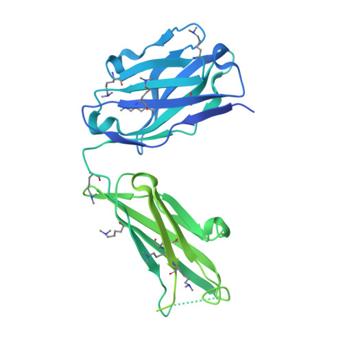
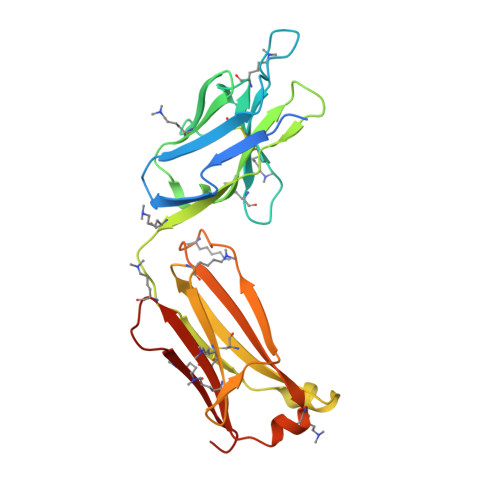
8HRH

SN-131/1B2 anti-MUC1 antibody with a glycopeptide


Experimental Data Snapshot

  • Method: X-RAY DIFFRACTION
  • Resolution: 2.07 Å
  • R-Value Free: 0.227 
  • R-Value Work: 0.192 

Starting Model: experimental
View more details

wwPDB Validation   3D Report Full Report


Ligand Structure Quality Assessment 


This is version 1.0 of the entry. See complete history


Literature

Structural and molecular insight into antibody recognition of dynamic neoepitopes in membrane tethered MUC1 of pancreatic cancer cells and secreted exosomes.

Wakui, H.Yokoi, Y.Horidome, C.Ose, T.Yao, M.Tanaka, Y.Hinou, H.Nishimura, S.I.

(2023) RSC Chem Biol 4: 564-572

  • DOI: https://doi.org/10.1039/d3cb00036b
  • Primary Citation of Related Structures:  
    8HRH

  • PubMed Abstract: 

    Pancreatic cancer is highly metastatic and has poor prognosis, mainly due to delayed detection, often after metastasis has occurred. A novel method to enable early detection and disease intervention is strongly needed. Here we unveil for the first time that pancreatic cancer cells (PANC-1) and secreted exosomes express MUC1 bearing cancer-relevant dynamic epitopes recognized specifically by an anti-MUC1 antibody (SN-131), which binds specifically core 1 but not core 2 type O -glycans found in normal cells. Comprehensive assessment of the essential epitope for SN-131 indicates that PANC-1 cells produce dominantly MUC1 with aberrant O -glycoforms such as Tn, T, and sialyl T (ST) antigens. Importantly, SN-131 showed the highest affinity with MUC1 bearing ST antigen at the immunodominant DTR motif ( K D = 1.58 nM) independent of the glycosylation states of other Ser/Thr residues in the MUC1 tandem repeats. The X-ray structure revealed that SN-131 interacts directly with Neu5Ac and root GalNAc of the ST antigen in addition to the proximal peptide region. Our results demonstrate that targeting O -glycosylated "dynamic neoepitopes" found in the membrane-tethered MUC1 is a promising therapeutic strategy for improving the treatment outcome of patients with pancreatic cancer.


  • Organizational Affiliation

    Field of Drug Discovery Research, Faculty of Advanced Life Science, and Graduate School of Life Science, Hokkaido University N21 W11 Kita-ku Sapporo 001-0021 Japan shin@sci.hokudai.ac.jp.


Macromolecules
Find similar proteins by:  (by identity cutoff)  |  3D Structure
Entity ID: 1
MoleculeChains Sequence LengthOrganismDetailsImage
Heavy chain of SN-131/1B2 antibody Fab467Mus musculusMutation(s): 0 
Entity Groups  
Sequence Clusters30% Identity50% Identity70% Identity90% Identity95% Identity100% Identity
Sequence Annotations
Expand
  • Reference Sequence
Find similar proteins by:  (by identity cutoff)  |  3D Structure
Entity ID: 2
MoleculeChains Sequence LengthOrganismDetailsImage
Light chain of SN-131/1B2 antibody Fab238Mus musculusMutation(s): 0 
Entity Groups  
Sequence Clusters30% Identity50% Identity70% Identity90% Identity95% Identity100% Identity
Sequence Annotations
Expand
  • Reference Sequence
Oligosaccharides

Help

Entity ID: 3
MoleculeChains Length2D Diagram Glycosylation3D Interactions
N-acetyl-alpha-neuraminic acid-(2-3)-beta-D-galactopyranose-(1-3)-2-acetamido-2-deoxy-alpha-D-galactopyranoseC [auth D]3O-Glycosylation
Glycosylation Resources
GlyTouCan:  G65562ZE
GlyCosmos:  G65562ZE
GlyGen:  G65562ZE
Small Molecules
Ligands 10 Unique
IDChains Name / Formula / InChI Key2D Diagram3D Interactions
ARG (Subject of Investigation/LOI)
Query on ARG

Download Ideal Coordinates CCD File 
G [auth A]ARGININE
C6 H15 N4 O2
ODKSFYDXXFIFQN-BYPYZUCNSA-O
N7P (Subject of Investigation/LOI)
Query on N7P

Download Ideal Coordinates CCD File 
D [auth A]1-ACETYL-L-PROLINE
C7 H11 N O3
GNMSLDIYJOSUSW-LURJTMIESA-N
ASP (Subject of Investigation/LOI)
Query on ASP

Download Ideal Coordinates CCD File 
E [auth A]ASPARTIC ACID
C4 H7 N O4
CKLJMWTZIZZHCS-REOHCLBHSA-N
TRS
Query on TRS

Download Ideal Coordinates CCD File 
I [auth A]2-AMINO-2-HYDROXYMETHYL-PROPANE-1,3-DIOL
C4 H12 N O3
LENZDBCJOHFCAS-UHFFFAOYSA-O
THR (Subject of Investigation/LOI)
Query on THR

Download Ideal Coordinates CCD File 
F [auth A]THREONINE
C4 H9 N O3
AYFVYJQAPQTCCC-GBXIJSLDSA-N
PRO (Subject of Investigation/LOI)
Query on PRO

Download Ideal Coordinates CCD File 
N [auth B]PROLINE
C5 H9 N O2
ONIBWKKTOPOVIA-BYPYZUCNSA-N
LPD
Query on LPD

Download Ideal Coordinates CCD File 
O [auth B]L-PROLINAMIDE
C5 H10 N2 O
VLJNHYLEOZPXFW-BYPYZUCNSA-N
PEG
Query on PEG

Download Ideal Coordinates CCD File 
J [auth A]
K [auth A]
P [auth B]
Q [auth B]
R [auth B]
J [auth A],
K [auth A],
P [auth B],
Q [auth B],
R [auth B],
S [auth B],
T [auth B]
DI(HYDROXYETHYL)ETHER
C4 H10 O3
MTHSVFCYNBDYFN-UHFFFAOYSA-N
GOL
Query on GOL

Download Ideal Coordinates CCD File 
L [auth A],
M [auth A]
GLYCEROL
C3 H8 O3
PEDCQBHIVMGVHV-UHFFFAOYSA-N
ALA (Subject of Investigation/LOI)
Query on ALA

Download Ideal Coordinates CCD File 
H [auth A]ALANINE
C3 H7 N O2
QNAYBMKLOCPYGJ-REOHCLBHSA-N
Modified Residues  1 Unique
IDChains TypeFormula2D DiagramParent
MLY
Query on MLY
A
L-PEPTIDE LINKINGC8 H18 N2 O2LYS
Experimental Data & Validation

Experimental Data

  • Method: X-RAY DIFFRACTION
  • Resolution: 2.07 Å
  • R-Value Free: 0.227 
  • R-Value Work: 0.192 
  • Space Group: P 65 2 2
Unit Cell:
Length ( Å )Angle ( ˚ )
a = 84.59α = 90
b = 84.59β = 90
c = 271.27γ = 120
Software Package:
Software NamePurpose
PHENIXrefinement
XDSdata scaling
Cootmodel building
PHASERphasing
XDSdata reduction

Structure Validation

View Full Validation Report


Ligand Structure Quality Assessment 


Entry History & Funding Information

Deposition Data


Funding OrganizationLocationGrant Number
Japan Society for the Promotion of Science (JSPS)Japan19H00918

Revision History  (Full details and data files)

  • Version 1.0: 2023-08-30
    Type: Initial release