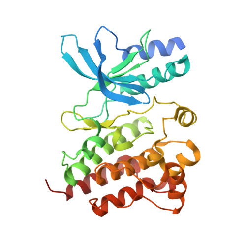
8OUU

Crystal structure of D1228V c-MET bound by compound 29


Experimental Data Snapshot

  • Method: X-RAY DIFFRACTION
  • Resolution: 1.77 Å
  • R-Value Free: 0.225 
  • R-Value Work: 0.202 
  • R-Value Observed: 0.203 

Starting Model: experimental
View more details

wwPDB Validation   3D Report Full Report


Ligand Structure Quality Assessment 


This is version 1.2 of the entry. See complete history


Literature

Discovery and Optimization of the First ATP Competitive Type-III c-MET Inhibitor.

Michaelides, I.N.Collie, G.W.Borjesson, U.Vasalou, C.Alkhatib, O.Barlind, L.Cheung, T.Dale, I.L.Embrey, K.J.Hennessy, E.J.Khurana, P.Koh, C.M.Lamb, M.L.Liu, J.Moss, T.A.O'Neill, D.J.Phillips, C.Shaw, J.Snijder, A.Storer, R.I.Stubbs, C.J.Han, F.Li, C.Qiao, J.Sun, D.Q.Wang, J.Wang, P.Yang, W.

(2023) J Med Chem 66: 8782-8807

  • DOI: https://doi.org/10.1021/acs.jmedchem.3c00401
  • Primary Citation of Related Structures:  
    8OUU, 8OUV, 8OV7, 8OVZ, 8OW3, 8OWG

  • PubMed Abstract: 

    Recent clinical reports have highlighted the need for wild-type (WT) and mutant dual inhibitors of c-MET kinase for the treatment of cancer. We report herein a novel chemical series of ATP competitive type-III inhibitors of WT and D1228V mutant c-MET. Using a combination of structure-based drug design and computational analyses, ligand 2 was optimized to a highly selective chemical series with nanomolar activities in biochemical and cellular settings. Representatives of the series demonstrate excellent pharmacokinetic profiles in rat in vivo studies with promising free-brain exposures, paving the way for the design of brain permeable drugs for the treatment of c-MET driven cancers.


  • Organizational Affiliation

    Discovery Sciences, R&D, AstraZeneca, Cambridge CB4 0WG, United Kingdom.


Macromolecules
Find similar proteins by:  (by identity cutoff)  |  3D Structure
Entity ID: 1
MoleculeChains Sequence LengthOrganismDetailsImage
Hepatocyte growth factor receptor
A, B
309Homo sapiensMutation(s): 1 
Gene Names: MET
EC: 2.7.10.1
UniProt & NIH Common Fund Data Resources
Find proteins for P08581 (Homo sapiens)
Explore P08581 
Go to UniProtKB:  P08581
PHAROS:  P08581
GTEx:  ENSG00000105976 
Entity Groups  
Sequence Clusters30% Identity50% Identity70% Identity90% Identity95% Identity100% Identity
UniProt GroupP08581
Sequence Annotations
Expand
  • Reference Sequence
Small Molecules
Ligands 4 Unique
IDChains Name / Formula / InChI Key2D Diagram3D Interactions
W49 (Subject of Investigation/LOI)
Query on W49

Download Ideal Coordinates CCD File 
F [auth A],
K [auth B]
5-(3-ethynyl-5-fluoranyl-1H-indazol-7-yl)-1-[(1S)-1-phenylethyl]pyrimidine-2,4-dione
C21 H15 F N4 O2
XZWWEKACMFDCHL-LBPRGKRZSA-N
GOL
Query on GOL

Download Ideal Coordinates CCD File 
C [auth A]GLYCEROL
C3 H8 O3
PEDCQBHIVMGVHV-UHFFFAOYSA-N
EDO
Query on EDO

Download Ideal Coordinates CCD File 
D [auth A],
G [auth B],
H [auth B]
1,2-ETHANEDIOL
C2 H6 O2
LYCAIKOWRPUZTN-UHFFFAOYSA-N
FMT
Query on FMT

Download Ideal Coordinates CCD File 
E [auth A],
I [auth B],
J [auth B]
FORMIC ACID
C H2 O2
BDAGIHXWWSANSR-UHFFFAOYSA-N
Experimental Data & Validation

Experimental Data

  • Method: X-RAY DIFFRACTION
  • Resolution: 1.77 Å
  • R-Value Free: 0.225 
  • R-Value Work: 0.202 
  • R-Value Observed: 0.203 
  • Space Group: P 21 21 21
Unit Cell:
Length ( Å )Angle ( ˚ )
a = 53.626α = 90
b = 56.738β = 90
c = 214.771γ = 90
Software Package:
Software NamePurpose
BUSTERrefinement
DIALSdata reduction
DIALSdata scaling
PHASERphasing

Structure Validation

View Full Validation Report


Ligand Structure Quality Assessment 


Entry History & Funding Information

Deposition Data

  • Released Date: 2023-07-05 
  • Deposition Author(s): Collie, G.W.

Funding OrganizationLocationGrant Number
Not funded--

Revision History  (Full details and data files)

  • Version 1.0: 2023-07-05
    Type: Initial release
  • Version 1.1: 2023-07-26
    Changes: Database references
  • Version 1.2: 2024-06-19
    Changes: Data collection