8P0D

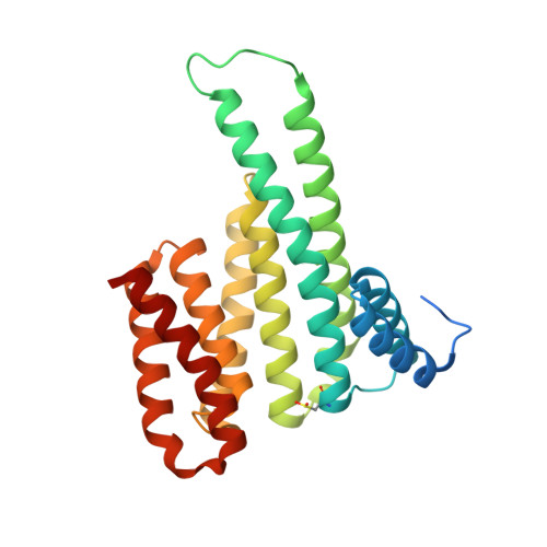
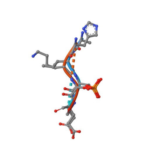
Human 14-3-3 sigma in complex with human MDM2 peptide


Experimental Data Snapshot

  • Method: X-RAY DIFFRACTION
  • Resolution: 1.31 Å
  • R-Value Free: 0.204 
  • R-Value Work: 0.177 
  • R-Value Observed: 0.179 

Starting Model: experimental
View more details

wwPDB Validation   3D Report Full Report


This is version 1.1 of the entry. See complete history


Literature

Characterizing the protein-protein interaction between MDM2 and 14-3-3 sigma ; proof of concept for small molecule stabilization.

Ward, J.A.Romartinez-Alonso, B.Kay, D.F.Bellamy-Carter, J.Thurairajah, B.Basran, J.Kwon, H.Leney, A.C.Macip, S.Roversi, P.Muskett, F.W.Doveston, R.G.

(2024) J Biol Chem 300: 105651-105651

  • DOI: https://doi.org/10.1016/j.jbc.2024.105651
  • Primary Citation of Related Structures:  
    8P0D

  • PubMed Abstract: 

    Mouse Double Minute 2 (MDM2) is a key negative regulator of the tumor suppressor protein p53. MDM2 overexpression occurs in many types of cancer and results in the suppression of WT p53. The 14-3-3 family of adaptor proteins are known to bind MDM2 and the 14-3-3σ isoform controls MDM2 cellular localization and stability to inhibit its activity. Therefore, small molecule stabilization of the 14-3-3σ/MDM2 protein-protein interaction (PPI) is a potential therapeutic strategy for the treatment of cancer. Here, we provide a detailed biophysical and structural characterization of the phosphorylation-dependent interaction between 14-3-3σ and peptides that mimic the 14-3-3 binding motifs within MDM2. The data show that di-phosphorylation of MDM2 at S166 and S186 is essential for high affinity 14-3-3 binding and that the binary complex formed involves one MDM2 di-phosphorylated peptide bound to a dimer of 14-3-3σ. However, the two phosphorylation sites do not simultaneously interact so as to bridge the 14-3-3 dimer in a 'multivalent' fashion. Instead, the two phosphorylated MDM2 motifs 'rock' between the two binding grooves of the dimer, which is unusual in the context of 14-3-3 proteins. In addition, we show that the 14-3-3σ-MDM2 interaction is amenable to small molecule stabilization. The natural product fusicoccin A forms a ternary complex with a 14-3-3σ dimer and an MDM2 di-phosphorylated peptide resulting in the stabilization of the 14-3-3σ/MDM2 PPI. This work serves as a proof-of-concept of the drugability of the 14-3-3/MDM2 PPI and paves the way toward the development of more selective and efficacious small molecule stabilizers.


  • Organizational Affiliation

    Leicester Institute for Structural and Chemical Biology, University of Leicester, Leicester, UK; Mechanisms of Cancer and Aging Laboratory, Department of Molecular and Cell Biology, University of Leicester, Leicester, UK.


Macromolecules
Find similar proteins by:  (by identity cutoff)  |  3D Structure
Entity ID: 1
MoleculeChains Sequence LengthOrganismDetailsImage
14-3-3 protein sigma259Homo sapiensMutation(s): 0 
Gene Names: SFNHME1
UniProt & NIH Common Fund Data Resources
Find proteins for P31947 (Homo sapiens)
Explore P31947 
Go to UniProtKB:  P31947
PHAROS:  P31947
GTEx:  ENSG00000175793 
Entity Groups  
Sequence Clusters30% Identity50% Identity70% Identity90% Identity95% Identity100% Identity
UniProt GroupP31947
Sequence Annotations
Expand
  • Reference Sequence
Find similar proteins by:  (by identity cutoff)  |  3D Structure
Entity ID: 2
MoleculeChains Sequence LengthOrganismDetailsImage
E3 ubiquitin-protein ligase Mdm231Homo sapiensMutation(s): 0 
EC: 2.3.2.27
UniProt & NIH Common Fund Data Resources
Find proteins for Q00987 (Homo sapiens)
Explore Q00987 
Go to UniProtKB:  Q00987
PHAROS:  Q00987
GTEx:  ENSG00000135679 
Entity Groups  
Sequence Clusters30% Identity50% Identity70% Identity90% Identity95% Identity100% Identity
UniProt GroupQ00987
Sequence Annotations
Expand
  • Reference Sequence
Small Molecules
Modified Residues  2 Unique
IDChains TypeFormula2D DiagramParent
CSD
Query on CSD
A
L-PEPTIDE LINKINGC3 H7 N O4 SCYS
SEP
Query on SEP
B
L-PEPTIDE LINKINGC3 H8 N O6 PSER
Experimental Data & Validation

Experimental Data

  • Method: X-RAY DIFFRACTION
  • Resolution: 1.31 Å
  • R-Value Free: 0.204 
  • R-Value Work: 0.177 
  • R-Value Observed: 0.179 
  • Space Group: C 2 2 21
Unit Cell:
Length ( Å )Angle ( ˚ )
a = 82.588α = 90
b = 111.824β = 90
c = 62.551γ = 90
Software Package:
Software NamePurpose
BUSTERrefinement
xia2data reduction
DIALSdata scaling
BUSTERphasing

Structure Validation

View Full Validation Report


Entry History & Funding Information

Deposition Data


Funding OrganizationLocationGrant Number
Wellcome TrustUnited Kingdom204801/Z/16/Z
Engineering and Physical Sciences Research CouncilUnited KingdomEP/W015803/1
Biotechnology and Biological Sciences Research Council (BBSRC)United KingdomBB/S019456/1
Biotechnology and Biological Sciences Research Council (BBSRC)United KingdomBB/M01116X/1
Wellcome TrustUnited Kingdom214090/Z/18/Z

Revision History  (Full details and data files)

  • Version 1.0: 2024-02-21
    Type: Initial release
  • Version 1.1: 2024-10-16
    Changes: Structure summary