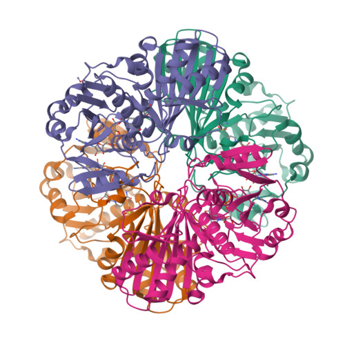
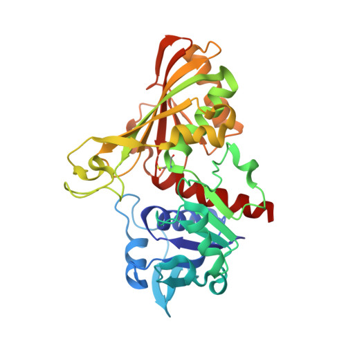
8P5F

Human wild-type GAPDH,orthorhombic form


Experimental Data Snapshot

  • Method: X-RAY DIFFRACTION
  • Resolution: 1.82 Å
  • R-Value Free: 0.189 
  • R-Value Work: 0.160 

Starting Model: experimental
View more details

wwPDB Validation   3D Report Full Report


Ligand Structure Quality Assessment 


This is version 1.2 of the entry. See complete history


Literature

S-nitrosylation and S-glutathionylation of GAPDH: Similarities, differences, and relationships.

Medvedeva, M.V.Kleimenov, S.Y.Samygina, V.R.Muronetz, V.I.Schmalhausen, E.V.

(2023) Biochim Biophys Acta Gen Subj 1867: 130418-130418

  • DOI: https://doi.org/10.1016/j.bbagen.2023.130418
  • Primary Citation of Related Structures:  
    8P5F

  • PubMed Abstract: 

    The aim of this work was to compare the effect of reversible post-translational modifications, S-nitrosylation and S-glutathionylation, on the properties of glyceraldehyde-3-phosphate dehydrogenase (GAPDH), and to reveal the mechanism of the relationship between these modifications. Comparison of S-nitrosylated and S-glutathionylated GAPDH showed that both modifications inactivate the enzyme and change its spatial structure, decreasing the thermal stability of the protein and increasing its sensitivity to trypsin cleavage. Both modifications are reversible in the presence of dithiothreitol, however, in the presence of reduced glutathione and glutaredoxin 1, the reactivation of S-glutathionylated GAPDH is much slower (10% in 2 h) compared to S-nitrosylated GAPDH (60% in 10 min). This suggests that S-glutathionylation is a much less reversible modification compared to S-nitrosylation. Incubation of HEK 293 T cells in the presence of H 2 O 2 or with the NO donor diethylamine NONOate results in accumulation of sulfenated GAPDH (by data of Western blotting) and S-glutathionylated GAPDH (by data of immunoprecipitation with anti-GSH antibodies). Besides GAPDH, a protein of 45 kDa was found to be sulfenated and S-glutathionylated in the cells treated with H 2 O 2 or NO. This protein was identified as beta-actin. The results of this study confirm the previously proposed hypothesis based on in vitro investigations, according to which S-nitrosylation of the catalytic cysteine residue (Cys152) of GAPDH with subsequent formation of cysteine sulfenic acid at Cys152 may promote its S-glutathionylation in the presence of cellular GSH. Presumably, the mechanism may be valid in the case of beta-actin.


  • Organizational Affiliation

    Faculty of Bioengineering and Bioinformatics, Lomonosov Moscow State University, Moscow 119991, Russia.


Macromolecules
Find similar proteins by:  (by identity cutoff)  |  3D Structure
Entity ID: 1
MoleculeChains Sequence LengthOrganismDetailsImage
Glyceraldehyde-3-phosphate dehydrogenaseA [auth AAA],
B [auth DDD],
C [auth EEE],
D [auth GGG]
335Homo sapiensMutation(s): 0 
Gene Names: GAPDHGAPDCDABP0047OK/SW-cl.12
EC: 1.2.1.12 (PDB Primary Data), 2.6.99 (PDB Primary Data)
UniProt & NIH Common Fund Data Resources
Find proteins for P04406 (Homo sapiens)
Explore P04406 
Go to UniProtKB:  P04406
PHAROS:  P04406
GTEx:  ENSG00000111640 
Entity Groups  
Sequence Clusters30% Identity50% Identity70% Identity90% Identity95% Identity100% Identity
UniProt GroupP04406
Sequence Annotations
Expand
  • Reference Sequence
Small Molecules
Ligands 3 Unique
IDChains Name / Formula / InChI Key2D Diagram3D Interactions
NAD (Subject of Investigation/LOI)
Query on NAD

Download Ideal Coordinates CCD File 
E [auth AAA],
F [auth DDD],
G [auth EEE],
K [auth GGG]
NICOTINAMIDE-ADENINE-DINUCLEOTIDE
C21 H27 N7 O14 P2
BAWFJGJZGIEFAR-NNYOXOHSSA-N
ZN
Query on ZN

Download Ideal Coordinates CCD File 
H [auth EEE],
J [auth EEE]
ZINC ION
Zn
PTFCDOFLOPIGGS-UHFFFAOYSA-N
ACT
Query on ACT

Download Ideal Coordinates CCD File 
I [auth EEE]ACETATE ION
C2 H3 O2
QTBSBXVTEAMEQO-UHFFFAOYSA-M
Modified Residues  1 Unique
IDChains TypeFormula2D DiagramParent
CSD
Query on CSD
A [auth AAA],
B [auth DDD],
C [auth EEE],
D [auth GGG]
L-PEPTIDE LINKINGC3 H7 N O4 SCYS
Experimental Data & Validation

Experimental Data

  • Method: X-RAY DIFFRACTION
  • Resolution: 1.82 Å
  • R-Value Free: 0.189 
  • R-Value Work: 0.160 
  • Space Group: P 21 21 21
Unit Cell:
Length ( Å )Angle ( ˚ )
a = 80.822α = 90
b = 132.812β = 90
c = 146.833γ = 90
Software Package:
Software NamePurpose
REFMACrefinement
XDSdata reduction
Aimlessdata scaling
MOLREPphasing

Structure Validation

View Full Validation Report


Ligand Structure Quality Assessment 


Entry History & Funding Information

Deposition Data


Funding OrganizationLocationGrant Number
Other governmentRussian Federation--

Revision History  (Full details and data files)

  • Version 1.0: 2023-07-05
    Type: Initial release
  • Version 1.1: 2023-07-12
    Changes: Database references
  • Version 1.2: 2024-11-06
    Changes: Data collection, Structure summary