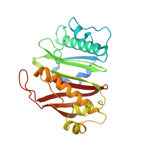
8QZI

Crystal structure of PptT-ACP from Mycobacterium tuberculosis


Experimental Data Snapshot

  • Method: X-RAY DIFFRACTION
  • Resolution: 2.50 Å
  • R-Value Free: 0.241 
  • R-Value Work: 0.196 
  • R-Value Observed: 0.198 

Starting Model: experimental
View more details

wwPDB Validation   3D Report Full Report


Ligand Structure Quality Assessment 


This is version 1.1 of the entry. See complete history


Literature

Catalytic Cycle of Type II 4'-Phosphopantetheinyl Transferases

Gavalda, S.Faille, A.Fioccola, S.Nguyen, M.C.Carivenc, C.Rottier, K.Rufin, Y.Saitta, S.Czaplicki, G.Guilhot, C.Chalut, C.Brut, M.Mourey, L.Pedelacq, J.D.

(2024) ACS Catal 14: 8561-8575

Macromolecules
Find similar proteins by:  (by identity cutoff)  |  3D Structure
Entity ID: 1
MoleculeChains Sequence LengthOrganismDetailsImage
4'-phosphopantetheinyl transferase PptT
A, B, C, D
247Mycobacterium tuberculosisMutation(s): 0 
Gene Names: pptT
EC: 2.7.8.7
UniProt
Find proteins for O33336 (Mycobacterium tuberculosis (strain ATCC 25618 / H37Rv))
Explore O33336 
Go to UniProtKB:  O33336
Entity Groups  
Sequence Clusters30% Identity50% Identity70% Identity90% Identity95% Identity100% Identity
UniProt GroupO33336
Sequence Annotations
Expand
  • Reference Sequence
Find similar proteins by:  (by identity cutoff)  |  3D Structure
Entity ID: 2
MoleculeChains Sequence LengthOrganismDetailsImage
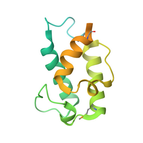
Phenolphthiocerol/phthiocerol polyketide synthase subunit C
E, F
172Mycobacterium tuberculosis H37RvMutation(s): 0 
Gene Names: ppsCRv2933
EC: 2.3.1.292
UniProt
Find proteins for P96202 (Mycobacterium tuberculosis (strain ATCC 25618 / H37Rv))
Explore P96202 
Go to UniProtKB:  P96202
Entity Groups  
Sequence Clusters30% Identity50% Identity70% Identity90% Identity95% Identity100% Identity
UniProt GroupP96202
Sequence Annotations
Expand
  • Reference Sequence
Small Molecules
Ligands 3 Unique
IDChains Name / Formula / InChI Key2D Diagram3D Interactions
COA (Subject of Investigation/LOI)
Query on COA

Download Ideal Coordinates CCD File 
G [auth A],
J [auth B],
M [auth C],
Q [auth D]
COENZYME A
C21 H36 N7 O16 P3 S
RGJOEKWQDUBAIZ-IBOSZNHHSA-N
IPA (Subject of Investigation/LOI)
Query on IPA

Download Ideal Coordinates CCD File 
P [auth C],
T [auth E]
ISOPROPYL ALCOHOL
C3 H8 O
KFZMGEQAYNKOFK-UHFFFAOYSA-N
MN (Subject of Investigation/LOI)
Query on MN

Download Ideal Coordinates CCD File 
H [auth A]
I [auth A]
K [auth B]
L [auth B]
N [auth C]
H [auth A],
I [auth A],
K [auth B],
L [auth B],
N [auth C],
O [auth C],
R [auth D],
S [auth D]
MANGANESE (II) ION
Mn
WAEMQWOKJMHJLA-UHFFFAOYSA-N
Modified Residues  1 Unique
IDChains TypeFormula2D DiagramParent
MSE
Query on MSE
E, F
L-PEPTIDE LINKINGC5 H11 N O2 SeMET
Experimental Data & Validation

Experimental Data

  • Method: X-RAY DIFFRACTION
  • Resolution: 2.50 Å
  • R-Value Free: 0.241 
  • R-Value Work: 0.196 
  • R-Value Observed: 0.198 
  • Space Group: P 1 21 1
Unit Cell:
Length ( Å )Angle ( ˚ )
a = 47.335α = 90
b = 280.843β = 92.829
c = 47.34γ = 90
Software Package:
Software NamePurpose
autoPROCdata processing
XDSdata reduction
Cootmodel building
PHASERphasing
PHENIXrefinement

Structure Validation

View Full Validation Report


Ligand Structure Quality Assessment 


Entry History & Funding Information

Deposition Data


Funding OrganizationLocationGrant Number
Agence Nationale de la Recherche (ANR)France09-BLAN-0298-01

Revision History  (Full details and data files)

  • Version 1.0: 2024-06-19
    Type: Initial release
  • Version 1.1: 2024-10-23
    Changes: Structure summary