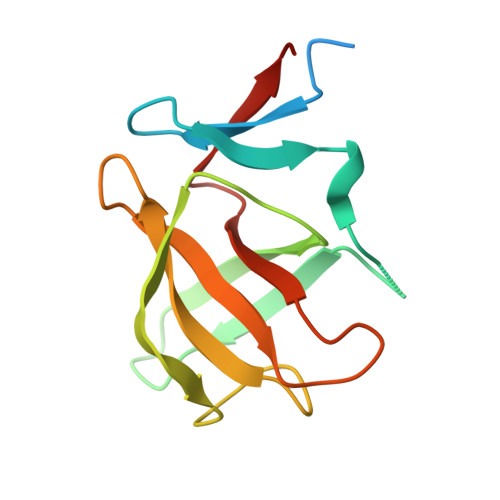
8RDP

Cereblon isoform 4 from Magnetospirillum gryphiswaldense in complex with spiro-isoxazol based compound 8a


Experimental Data Snapshot

  • Method: X-RAY DIFFRACTION
  • Resolution: 1.76 Å
  • R-Value Free: 0.230 
  • R-Value Work: 0.186 
  • R-Value Observed: 0.188 

Starting Model: experimental
View more details

wwPDB Validation   3D Report Full Report


Ligand Structure Quality Assessment 


This is version 1.0 of the entry. See complete history


Literature

Discovery and characterization of potent spiro-isoxazole-based cereblon ligands with a novel binding mode.

Shevalev, R.Bischof, L.Sapegin, A.Bunev, A.Olga, G.Kantin, G.Kalinin, S.Hartmann, M.D.

(2024) Eur J Med Chem 270: 116328-116328

  • DOI: https://doi.org/10.1016/j.ejmech.2024.116328
  • Primary Citation of Related Structures:  
    8RDP, 8RDQ, 8RDR, 8RDS, 8RDT

  • PubMed Abstract: 

    The vast majority of current cereblon (CRBN) ligands is based on the thalidomide scaffold, relying on glutarimide as the core binding moiety. With this architecture, most of these ligands inherit the overall binding mode, interactions with neo-substrates, and thereby potentially also the cytotoxic and teratogenic properties of the parent thalidomide. In this work, by incorporating a spiro-linker to the glutarimide moiety, we have generated a new chemotype that exhibits an unprecedented binding mode for glutarimide-based CRBN ligands. In total, 16 spirocyclic glutarimide derivatives incorporating an isoxazole moiety were synthesized and tested for different criteria. In particular, all ligands showed a favorable lipophilicity, and several were able to outperform the binding affinity of thalidomide as a reference. In addition, all compounds showed favorable cytotoxicity profiles in myeloma cell lines and human peripheral blood mononuclear cells. The novel binding mode, which we determined in co-crystal structures, provides explanations for these improved properties: The incorporation of the spiro-isoxazole changes both the conformation of the glutarimide moiety within the canonical tri-trp pocket and the orientation of the protruding moiety. In this new orientation it forms additional hydrophobic interactions and is not available for direct interactions with the canonical neo-substrates. We therefore propose this chemotype as an attractive building block for the design of PROTACs.


  • Organizational Affiliation

    Institute of Chemistry, Saint Petersburg State University, Saint Petersburg, Russia.


Macromolecules
Find similar proteins by:  (by identity cutoff)  |  3D Structure
Entity ID: 1
MoleculeChains Sequence LengthOrganismDetailsImage
Cereblon isoform 4
A, B, C
125Magnetospirillum gryphiswaldenseMutation(s): 0 
Gene Names: MGR_0879
UniProt
Find proteins for A4TVL0 (Magnetospirillum gryphiswaldense)
Explore A4TVL0 
Go to UniProtKB:  A4TVL0
Entity Groups  
Sequence Clusters30% Identity50% Identity70% Identity90% Identity95% Identity100% Identity
UniProt GroupA4TVL0
Sequence Annotations
Expand
  • Reference Sequence
Small Molecules
Ligands 4 Unique
IDChains Name / Formula / InChI Key2D Diagram3D Interactions
A1HZ7 (Subject of Investigation/LOI)
Query on A1HZ7

Download Ideal Coordinates CCD File 
E [auth A],
H [auth B],
K [auth C]
(5~{S})-3-(2-chlorophenyl)-1-oxa-2,9-diazaspiro[4.5]dec-2-ene-8,10-dione
C13 H11 Cl N2 O3
BUCCPXBJMQOJMQ-ZDUSSCGKSA-N
PO4
Query on PO4

Download Ideal Coordinates CCD File 
I [auth B]PHOSPHATE ION
O4 P
NBIIXXVUZAFLBC-UHFFFAOYSA-K
ZN
Query on ZN

Download Ideal Coordinates CCD File 
D [auth A],
G [auth B],
J [auth C]
ZINC ION
Zn
PTFCDOFLOPIGGS-UHFFFAOYSA-N
NA
Query on NA

Download Ideal Coordinates CCD File 
F [auth A]SODIUM ION
Na
FKNQFGJONOIPTF-UHFFFAOYSA-N
Experimental Data & Validation

Experimental Data

  • Method: X-RAY DIFFRACTION
  • Resolution: 1.76 Å
  • R-Value Free: 0.230 
  • R-Value Work: 0.186 
  • R-Value Observed: 0.188 
  • Space Group: P 21 21 21
Unit Cell:
Length ( Å )Angle ( ˚ )
a = 56.356α = 90
b = 59.597β = 90
c = 87.875γ = 90
Software Package:
Software NamePurpose
REFMACrefinement
XDSdata reduction
Cootmodel building

Structure Validation

View Full Validation Report


Ligand Structure Quality Assessment 


Entry History & Funding Information

Deposition Data


Funding OrganizationLocationGrant Number
Max Planck SocietyGermany--

Revision History  (Full details and data files)

  • Version 1.0: 2024-04-10
    Type: Initial release