8TN6

De novo designed protein binds poly ADP ribose polymerase inhibitors (PARPi) - holo rucaparib


Experimental Data Snapshot

  • Method: X-RAY DIFFRACTION
  • Resolution: 1.36 Å
  • R-Value Free: 0.217 
  • R-Value Work: 0.195 
  • R-Value Observed: 0.196 

wwPDB Validation   3D Report Full Report


Ligand Structure Quality Assessment 


This is version 1.0 of the entry. See complete history


Literature

De novo design of drug-binding proteins with predictable binding energy and specificity.

Lu, L.Gou, X.Tan, S.K.Mann, S.I.Yang, H.Zhong, X.Gazgalis, D.Valdiviezo, J.Jo, H.Wu, Y.Diolaiti, M.E.Ashworth, A.Polizzi, N.F.DeGrado, W.F.

(2024) Science 384: 106-112

  • DOI: https://doi.org/10.1126/science.adl5364
  • Primary Citation of Related Structures:  
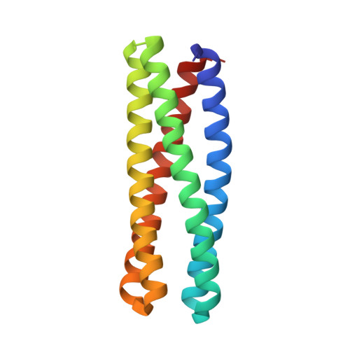
    8TN1, 8TN6, 8TNB, 8TNC, 8TND

  • PubMed Abstract: 

    The de novo design of small molecule-binding proteins has seen exciting recent progress; however, high-affinity binding and tunable specificity typically require laborious screening and optimization after computational design. We developed a computational procedure to design a protein that recognizes a common pharmacophore in a series of poly(ADP-ribose) polymerase-1 inhibitors. One of three designed proteins bound different inhibitors with affinities ranging from <5 nM to low micromolar. X-ray crystal structures confirmed the accuracy of the designed protein-drug interactions. Molecular dynamics simulations informed the role of water in binding. Binding free energy calculations performed directly on the designed models were in excellent agreement with the experimentally measured affinities. We conclude that de novo design of high-affinity small molecule-binding proteins with tuned interaction energies is feasible entirely from computation.


  • Organizational Affiliation

    Department of Pharmaceutical Chemistry & Cardiovascular Research Institute, University of California, San Francisco, CA 94158, USA.


Macromolecules
Find similar proteins by:  (by identity cutoff)  |  3D Structure
Entity ID: 1
MoleculeChains Sequence LengthOrganismDetailsImage
De novo designed protein
A, B, C
147synthetic constructMutation(s): 0 
Entity Groups  
Sequence Clusters30% Identity50% Identity70% Identity90% Identity95% Identity100% Identity
Sequence Annotations
Expand
  • Reference Sequence
Experimental Data & Validation

Experimental Data

  • Method: X-RAY DIFFRACTION
  • Resolution: 1.36 Å
  • R-Value Free: 0.217 
  • R-Value Work: 0.195 
  • R-Value Observed: 0.196 
  • Space Group: P 61 2 2
Unit Cell:
Length ( Å )Angle ( ˚ )
a = 91.252α = 90
b = 91.252β = 90
c = 201.951γ = 120
Software Package:
Software NamePurpose
PHENIXrefinement
XDSdata reduction
pointlessdata scaling
PHENIXphasing
PHENIXrefinement
Cootmodel building

Structure Validation

View Full Validation Report


Ligand Structure Quality Assessment 


Entry History & Funding Information

Deposition Data


Funding OrganizationLocationGrant Number
National Institutes of Health/National Institute of General Medical Sciences (NIH/NIGMS)United States2R35GM122603

Revision History  (Full details and data files)

  • Version 1.0: 2024-04-24
    Type: Initial release