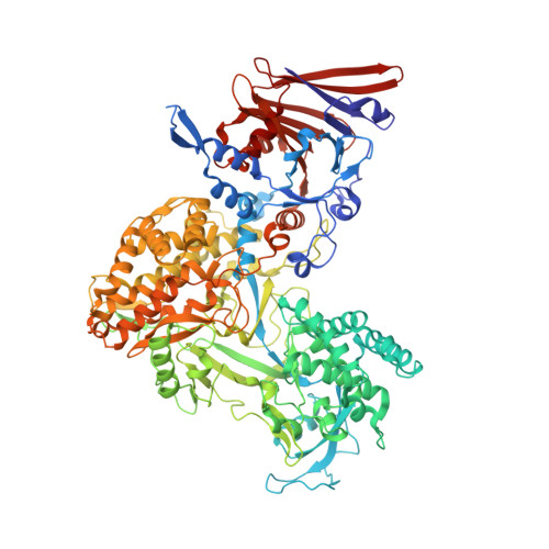
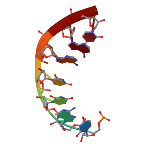
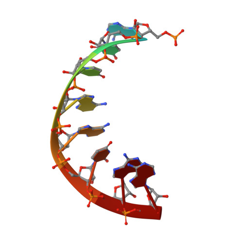
8W1O

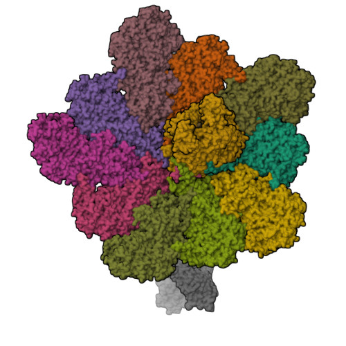
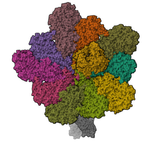
Cryo-EM structure of BTV virion


Experimental Data Snapshot

  • Method: ELECTRON MICROSCOPY
  • Resolution: 2.80 Å
  • Aggregation State: PARTICLE 
  • Reconstruction Method: SINGLE PARTICLE 

wwPDB Validation   3D Report Full Report


This is version 1.1 of the entry. See complete history


Literature

RNA genome packaging and capsid assembly of bluetongue virus visualized in host cells.

Xia, X.Sung, P.Y.Martynowycz, M.W.Gonen, T.Roy, P.Zhou, Z.H.

(2024) Cell 187: 2236-2249.e17

  • DOI: https://doi.org/10.1016/j.cell.2024.03.007
  • Primary Citation of Related Structures:  
    8W12, 8W19, 8W1C, 8W1I, 8W1O, 8W1R, 8W1S

  • PubMed Abstract: 

    Unlike those of double-stranded DNA (dsDNA), single-stranded DNA (ssDNA), and ssRNA viruses, the mechanism of genome packaging of dsRNA viruses is poorly understood. Here, we combined the techniques of high-resolution cryoelectron microscopy (cryo-EM), cellular cryoelectron tomography (cryo-ET), and structure-guided mutagenesis to investigate genome packaging and capsid assembly of bluetongue virus (BTV), a member of the Reoviridae family of dsRNA viruses. A total of eleven assembly states of BTV capsid were captured, with resolutions up to 2.8 Å, with most visualized in the host cytoplasm. ATPase VP6 was found underneath the vertices of capsid shell protein VP3 as an RNA-harboring pentamer, facilitating RNA packaging. RNA packaging expands the VP3 shell, which then engages middle- and outer-layer proteins to generate infectious virions. These revealed "duality" characteristics of the BTV assembly mechanism reconcile previous contradictory co-assembly and core-filling models and provide insights into the mysterious RNA packaging and capsid assembly of Reoviridae members and beyond.


  • Organizational Affiliation

    Department of Microbiology, Immunology and Molecular Genetics, University of California, Los Angeles, Los Angeles, CA 90095, USA; California NanoSystems Institute, University of California, Los Angeles, Los Angeles, CA 90095, USA.


Macromolecules

Find similar proteins by:  (by identity cutoff)  |  3D Structure
Entity ID: 1
MoleculeChains Sequence LengthOrganismDetailsImage
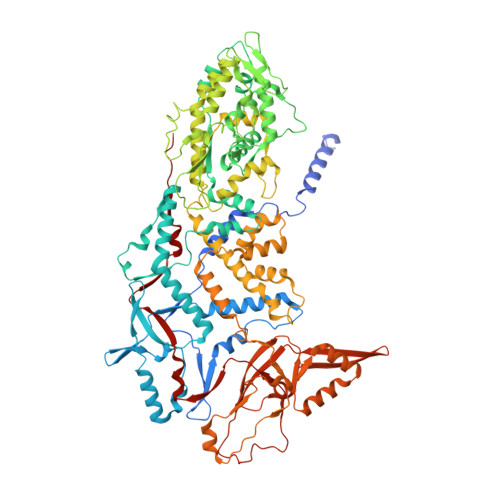
Core protein VP3
A, B, C, D, E
A, B, C, D, E, F, G, H, I, J
901Bluetongue virus (serotype 1 / isolate South Africa)Mutation(s): 0 
UniProt
Find proteins for P56582 (Bluetongue virus 1 (isolate South Africa))
Explore P56582 
Go to UniProtKB:  P56582
Entity Groups  
Sequence Clusters30% Identity50% Identity70% Identity90% Identity95% Identity100% Identity
UniProt GroupP56582
Sequence Annotations
Expand
  • Reference Sequence
Find similar proteins by:  (by identity cutoff)  |  3D Structure
Entity ID: 2
MoleculeChains Sequence LengthOrganismDetailsImage
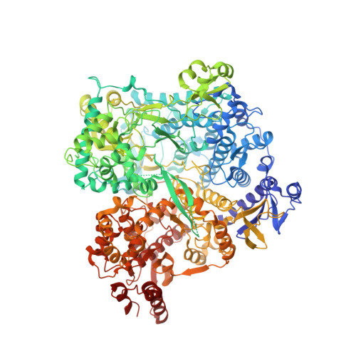
RNA-directed RNA polymerase1,302Bluetongue virus (serotype 1 / isolate South Africa)Mutation(s): 0 
EC: 2.7.7.48
UniProt
Find proteins for W0G557 (Bluetongue virus 1)
Explore W0G557 
Go to UniProtKB:  W0G557
Entity Groups  
Sequence Clusters30% Identity50% Identity70% Identity90% Identity95% Identity100% Identity
UniProt GroupW0G557
Sequence Annotations
Expand
  • Reference Sequence
Find similar proteins by:  (by identity cutoff)  |  3D Structure
Entity ID: 5
MoleculeChains Sequence LengthOrganismDetailsImage
Outer capsid protein VP2961Bluetongue virus (serotype 1 / isolate South Africa)Mutation(s): 0 
UniProt
Find proteins for B7U676 (Bluetongue virus 1)
Explore B7U676 
Go to UniProtKB:  B7U676
Entity Groups  
Sequence Clusters30% Identity50% Identity70% Identity90% Identity95% Identity100% Identity
UniProt GroupB7U676
Sequence Annotations
Expand
  • Reference Sequence

Find similar nucleic acids by:  Sequence   |   3D Structure  

Entity ID: 3
MoleculeChains LengthOrganismImage
RNA-17Bluetongue virus (serotype 1 / isolate South Africa)
Sequence Annotations
Expand
  • Reference Sequence

Find similar nucleic acids by:  Sequence   |   3D Structure  

Entity ID: 4
MoleculeChains LengthOrganismImage
RNA-29Bluetongue virus (serotype 1 / isolate South Africa)
Sequence Annotations
Expand
  • Reference Sequence
Experimental Data & Validation

Experimental Data

  • Method: ELECTRON MICROSCOPY
  • Resolution: 2.80 Å
  • Aggregation State: PARTICLE 
  • Reconstruction Method: SINGLE PARTICLE 
EM Software:
TaskSoftware PackageVersion
RECONSTRUCTIONRELION3.1
MODEL REFINEMENTPHENIX1.19.2

Structure Validation

View Full Validation Report


Entry History & Funding Information

Deposition Data


Funding OrganizationLocationGrant Number
National Institutes of Health/National Institute Of Allergy and Infectious Diseases (NIH/NIAID)United StatesAI094386
Wellcome TrustUnited KingdomWT221749/20/Z

Revision History  (Full details and data files)

  • Version 1.0: 2024-04-24
    Type: Initial release
  • Version 1.1: 2024-05-08
    Changes: Database references