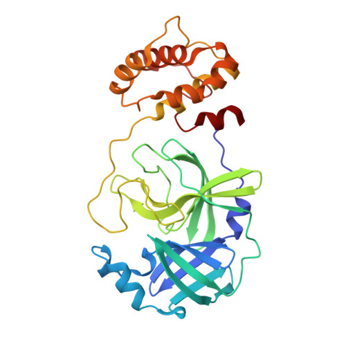
8WUR

Crystal structure of SARS-Cov-2 main protease D48N mutant in complex with shikonin


Experimental Data Snapshot

  • Method: X-RAY DIFFRACTION
  • Resolution: 2.08 Å
  • R-Value Free: 0.238 
  • R-Value Work: 0.211 
  • R-Value Observed: 0.212 

Starting Model: experimental
View more details

wwPDB Validation   3D Report Full Report


Ligand Structure Quality Assessment 


This is version 1.0 of the entry. See complete history


Literature

Structural Basis for the Inhibition of SARS-CoV-2 M pro D48N Mutant by Shikonin and PF-07321332.

Zhao, Z.Zhu, Q.Zhou, X.Li, W.Yin, X.Li, J.

(2023) Viruses 16

  • DOI: https://doi.org/10.3390/v16010065
  • Primary Citation of Related Structures:  
    7XB4, 8WUR

  • PubMed Abstract: 

    Preventing the spread of SARS-CoV-2 and its variants is crucial in the fight against COVID-19. Inhibition of the main protease (M pro ) of SARS-CoV-2 is the key to disrupting viral replication, making M pro a promising target for therapy. PF-07321332 and shikonin have been identified as effective broad-spectrum inhibitors of SARS-CoV-2 M pro . The crystal structures of SARS-CoV-2 M pro bound to PF-07321332 and shikonin have been resolved in previous studies. However, the exact mechanism regarding how SARS-CoV-2 M pro mutants impact their binding modes largely remains to be investigated. In this study, we expressed a SARS-CoV-2 M pro mutant, carrying the D48N substitution, representing a class of mutations located near the active sites of M pro . The crystal structures of M pro D48N in complex with PF-07321332 and shikonin were solved. A detailed analysis of the interactions between M pro D48N and two inhibitors provides key insights into the binding pattern and its structural determinants. Further, the binding patterns of the two inhibitors to M pro D48N mutant and wild-type M pro were compared in detail. This study illustrates the possible conformational changes when the M pro D48N mutant is bound to inhibitors. Structural insights derived from this study will inform the development of new drugs against novel coronaviruses.


  • Organizational Affiliation

    College of Pharmaceutical Sciences, Gannan Medical University, Ganzhou 341000, China.


Macromolecules
Find similar proteins by:  (by identity cutoff)  |  3D Structure
Entity ID: 1
MoleculeChains Sequence LengthOrganismDetailsImage
3C-like proteinase nsp5
A, B
296Severe acute respiratory syndrome coronavirus 2Mutation(s): 1 
EC: 3.4.22.69
UniProt
Find proteins for P0DTC1 (Severe acute respiratory syndrome coronavirus 2)
Explore P0DTC1 
Go to UniProtKB:  P0DTC1
Entity Groups  
Sequence Clusters30% Identity50% Identity70% Identity90% Identity95% Identity100% Identity
UniProt GroupP0DTC1
Sequence Annotations
Expand
  • Reference Sequence
Small Molecules
Ligands 1 Unique
IDChains Name / Formula / InChI Key2D Diagram3D Interactions
FNO (Subject of Investigation/LOI)
Query on FNO

Download Ideal Coordinates CCD File 
C [auth A]2-[(1R)-4-methyl-1-oxidanyl-pent-3-enyl]-5,8-bis(oxidanyl)naphthalene-1,4-dione
C16 H16 O5
NEZONWMXZKDMKF-SNVBAGLBSA-N
Experimental Data & Validation

Experimental Data

  • Method: X-RAY DIFFRACTION
  • Resolution: 2.08 Å
  • R-Value Free: 0.238 
  • R-Value Work: 0.211 
  • R-Value Observed: 0.212 
  • Space Group: P 21 21 21
Unit Cell:
Length ( Å )Angle ( ˚ )
a = 68.052α = 90
b = 102.944β = 90
c = 103.687γ = 90
Software Package:
Software NamePurpose
PHENIXrefinement
XDSdata reduction
XDSdata scaling
PHENIXphasing

Structure Validation

View Full Validation Report


Ligand Structure Quality Assessment 


Entry History & Funding Information

Deposition Data


Funding OrganizationLocationGrant Number
Not funded--

Revision History  (Full details and data files)

  • Version 1.0: 2024-11-13
    Type: Initial release