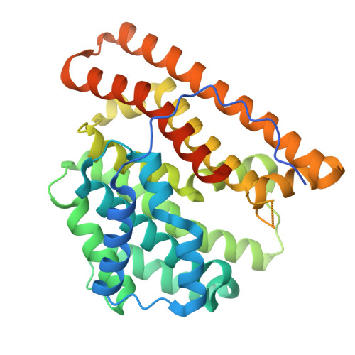
9C7J

Crystal structure of caryolan-1-ol synthase complexed with 2-fluorofarnesyl diphosphate


Experimental Data Snapshot

  • Method: X-RAY DIFFRACTION
  • Resolution: 2.65 Å
  • R-Value Free: 0.249 
  • R-Value Work: 0.204 
  • R-Value Observed: 0.206 

Starting Model: experimental
View more details

wwPDB Validation   3D Report Full Report


Ligand Structure Quality Assessment 


This is version 1.0 of the entry. See complete history


Literature

Crystal Structure of Caryolan-1-ol Synthase, a Sesquiterpene Synthase Catalyzing an Initial Anti-Markovnikov Cyclization Reaction.

Kumar, R.P.Matos, J.O.Black, B.Y.Ellenburg, W.H.Chen, J.Patterson, M.Gehtman, J.A.Theobald, D.L.Krauss, I.J.Oprian, D.D.

(2024) Biochemistry 

  • DOI: https://doi.org/10.1021/acs.biochem.4c00547
  • Primary Citation of Related Structures:  
    9C7I, 9C7J, 9C7K, 9C7L, 9C7M

  • PubMed Abstract: 

    In a continuing effort to understand reaction mechanisms of terpene synthases catalyzing initial anti-Markovnikov cyclization reactions, we solved the X-ray crystal structure of (+)-caryolan-1-ol synthase (CS) from Streptomyces griseus , with and without an inactive analog of the farnesyl diphosphate (FPP) substrate, 2-fluorofarnesyl diphosphate (2FFPP), bound in the active site of the enzyme. The CS-2FFPP structure was solved to 2.65 Å resolution and showed the ligand in an elongated orientation, incapable of undergoing the initial cyclization event to form a C1-C11 bond. Intriguingly, the apo CS structure (2.2 Å) also had electron density in the active site, in this case, well fit by a curled-up tetraethylene glycol molecule recruited, presumably, from the crystallization medium. The density was also well fit by a molecule of farnesene suggesting that the structure may mimic an intermediate along the reaction coordinate. The curled-up conformation of tetraethylene glycol was accompanied by dramatic rotation of some active-site residues in comparison to the 2FFPP-structure. Most notably, W56 and F183 undergo 90° rotations between the 2FFPP complex and apoenzyme structures, suggesting that these residues provide interactions that help curl the tetraethylene glycol molecule in the active site, and by extension perhaps also a derivative of the FPP substrate in the normal course of the cyclization reaction. In support of this proposal, the CS W56L and F183A variants were observed to be severely restricted in their ability to catalyze C1-C11 cyclization of the FPP substrate and instead produced predominantly acyclic terpene products dominated by farnesol, β-farnesene, and nerolidol.


  • Organizational Affiliation

    Department of Biochemistry, Brandeis University, Waltham, Massachusetts 02454, United States.


Macromolecules
Find similar proteins by:  (by identity cutoff)  |  3D Structure
Entity ID: 1
MoleculeChains Sequence LengthOrganismDetailsImage
(+)-caryolan-1-ol synthase
A, B, C, D
349Streptomyces griseusMutation(s): 0 
Gene Names: gcoASGR_2079
EC: 4.2.1.138 (PDB Primary Data), 4.2.3.89 (PDB Primary Data)
UniProt
Find proteins for B1W019 (Streptomyces griseus subsp. griseus (strain JCM 4626 / CBS 651.72 / NBRC 13350 / KCC S-0626 / ISP 5235))
Explore B1W019 
Go to UniProtKB:  B1W019
Entity Groups  
Sequence Clusters30% Identity50% Identity70% Identity90% Identity95% Identity100% Identity
UniProt GroupB1W019
Sequence Annotations
Expand
  • Reference Sequence
Small Molecules
Ligands 4 Unique
IDChains Name / Formula / InChI Key2D Diagram3D Interactions
FPF (Subject of Investigation/LOI)
Query on FPF

Download Ideal Coordinates CCD File 
E [auth A],
H [auth B],
K [auth C],
O [auth D]
(2Z,6E)-2-fluoro-3,7,11-trimethyldodeca-2,6,10-trien-1-yl trihydrogen diphosphate
C15 H27 F O7 P2
DXHJZCKJWKWTCH-WISUYLHISA-N
DPO
Query on DPO

Download Ideal Coordinates CCD File 
J [auth B]DIPHOSPHATE
O7 P2
XPPKVPWEQAFLFU-UHFFFAOYSA-J
PO4
Query on PO4

Download Ideal Coordinates CCD File 
F [auth A]
I [auth B]
L [auth C]
N [auth C]
P [auth D]
F [auth A],
I [auth B],
L [auth C],
N [auth C],
P [auth D],
Q [auth D]
PHOSPHATE ION
O4 P
NBIIXXVUZAFLBC-UHFFFAOYSA-K
MG (Subject of Investigation/LOI)
Query on MG

Download Ideal Coordinates CCD File 
G [auth A],
M [auth C],
R [auth D]
MAGNESIUM ION
Mg
JLVVSXFLKOJNIY-UHFFFAOYSA-N
Experimental Data & Validation

Experimental Data

  • Method: X-RAY DIFFRACTION
  • Resolution: 2.65 Å
  • R-Value Free: 0.249 
  • R-Value Work: 0.204 
  • R-Value Observed: 0.206 
  • Space Group: P 1 21 1
Unit Cell:
Length ( Å )Angle ( ˚ )
a = 68.648α = 90
b = 89.606β = 90.71
c = 114.814γ = 90
Software Package:
Software NamePurpose
PHENIXrefinement
SCALAdata scaling
XDSdata reduction
PHASERphasing

Structure Validation

View Full Validation Report


Ligand Structure Quality Assessment 


Entry History & Funding Information

Deposition Data


Funding OrganizationLocationGrant Number
Not funded--

Revision History  (Full details and data files)

  • Version 1.0: 2024-11-06
    Type: Initial release