Structural basis of hexose specificity and catalytic properties of Kluyveromyces lactis glucokinase KlGlk1
Weisse, R.H., Kettner, K., Lilie, H., Funk, L., Roedel, G., Tittmann, K., Strater, N., Kriegel, T.M.To be published.
Experimental Data Snapshot
Starting Model: experimental
View more details
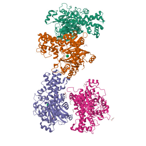
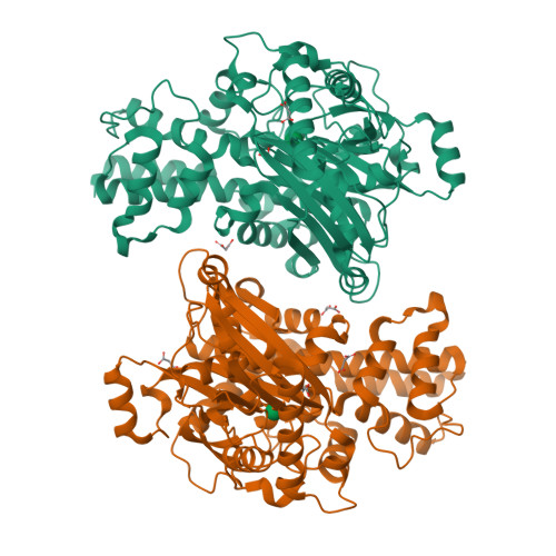
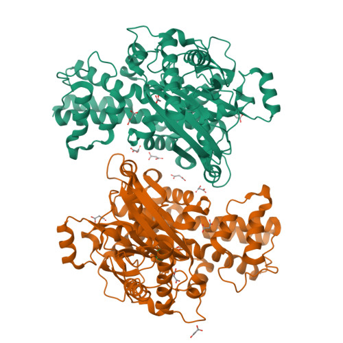
Entity ID: 1 | |||||
|---|---|---|---|---|---|
| Molecule | Chains | Sequence Length | Organism | Details | Image |
| Glucokinase-1 | 481 | Kluyveromyces lactis | Mutation(s): 1 Gene Names: GLK1, KLLA0C01155g EC: 2.7.1.2 (PDB Primary Data), 2.7.1.1 (PDB Primary Data) |  | |
UniProt | |||||
Find proteins for Q6CUZ3 (Kluyveromyces lactis (strain ATCC 8585 / CBS 2359 / DSM 70799 / NBRC 1267 / NRRL Y-1140 / WM37)) Explore Q6CUZ3 Go to UniProtKB: Q6CUZ3 | |||||
Entity Groups | |||||
| Sequence Clusters | 30% Identity50% Identity70% Identity90% Identity95% Identity100% Identity | ||||
| UniProt Group | Q6CUZ3 | ||||
Sequence AnnotationsExpand | |||||
| |||||
| Ligands 2 Unique | |||||
|---|---|---|---|---|---|
| ID | Chains | Name / Formula / InChI Key | 2D Diagram | 3D Interactions | |
| MAN (Subject of Investigation/LOI) Query on MAN | E [auth A], I [auth B], N [auth C], U [auth D] | alpha-D-mannopyranose C6 H12 O6 WQZGKKKJIJFFOK-PQMKYFCFSA-N |  | ||
| MLI Query on MLI | AA [auth D] F [auth A] G [auth A] H [auth A] J [auth B] | MALONATE ION C3 H2 O4 OFOBLEOULBTSOW-UHFFFAOYSA-L |  | ||
| Length ( Å ) | Angle ( ˚ ) |
|---|---|
| a = 202.412 | α = 90 |
| b = 88.34 | β = 89.92 |
| c = 212.45 | γ = 90 |
| Software Name | Purpose |
|---|---|
| PHENIX | refinement |
| DIALS | data reduction |
| Aimless | data scaling |
| Funding Organization | Location | Grant Number |
|---|---|---|
| Helmholtz Association | Germany | -- |