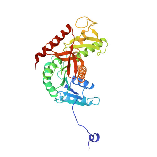
9LDT

DESIGN AND SYNTHESIS OF NEW ENZYMES BASED ON THE LACTATE DEHYDROGENASE FRAMEWORK


Experimental Data Snapshot

  • Method: X-RAY DIFFRACTION
  • Resolution: 2.00 Å
  • R-Value Observed: 0.233 

wwPDB Validation   3D Report Full Report


This is version 2.1 of the entry. See complete history


Literature

Design and synthesis of new enzymes based on the lactate dehydrogenase framework.

Dunn, C.R.Wilks, H.M.Halsall, D.J.Atkinson, T.Clarke, A.R.Muirhead, H.Holbrook, J.J.

(1991) Philos Trans R Soc London,ser B 332: 177-184

  • DOI: https://doi.org/10.1098/rstb.1991.0047
  • Primary Citation of Related Structures:  
    9LDB, 9LDT

  • PubMed Abstract: 

    Analysis of the mechanism and structure of lactate dehydrogenases is summarized in a map of the catalytic pathway. Chemical probes, single tryptophan residues inserted at specific sites and a crystal structure reveal slow movements of the protein framework that discriminate between closely related small substrates. Only small and correctly charged substrates allow the protein to engulf the substrate in an internal vacuole that is isolated from solvent protons, in which water is frozen and hydride transfer is rapid. The closed vacuole is very sensitive to the size and charge of the substrate and provides discrimination between small substrates that otherwise have too few functional groups to be distinguished at a solvated protein surface. This model was tested against its ability to successfully predict the design and synthesis of new enzymes such as L-hydroxyisocaproate dehydrogenase and fully active malate dehydrogenase. Solvent friction limits the rate of forming the vacuole and thus the maximum rate of catalysis.


  • Organizational Affiliation

    University of Bristol Molecular Recognition Centre, School of Medical Sciences, U.K.


Macromolecules
Find similar proteins by:  (by identity cutoff)  |  3D Structure
Entity ID: 1
MoleculeChains Sequence LengthOrganismDetailsImage
LACTATE DEHYDROGENASE
A, B
332Sus scrofaMutation(s): 0 
EC: 1.1.1.27
UniProt
Find proteins for P00339 (Sus scrofa)
Explore P00339 
Go to UniProtKB:  P00339
Entity Groups  
Sequence Clusters30% Identity50% Identity70% Identity90% Identity95% Identity100% Identity
UniProt GroupP00339
Sequence Annotations
Expand
  • Reference Sequence
Experimental Data & Validation

Experimental Data

  • Method: X-RAY DIFFRACTION
  • Resolution: 2.00 Å
  • R-Value Observed: 0.233 
  • Space Group: P 21 21 2
Unit Cell:
Length ( Å )Angle ( ˚ )
a = 60.3α = 90
b = 136.39β = 90
c = 86.05γ = 90
Software Package:
Software NamePurpose
PROLSQrefinement

Structure Validation

View Full Validation Report


Entry History 

Deposition Data

Revision History  (Full details and data files)

  • Version 1.0: 1993-10-31
    Type: Initial release
  • Version 1.1: 2008-03-25
    Changes: Version format compliance
  • Version 1.2: 2011-07-13
    Changes: Advisory, Derived calculations, Version format compliance
  • Version 1.3: 2017-11-29
    Changes: Derived calculations, Other
  • Version 2.0: 2023-09-27
    Type: Remediation
    Changes: Advisory, Atomic model, Data collection, Database references, Derived calculations, Other, Refinement description
  • Version 2.1: 2024-10-16
    Changes: Structure summary