1OD1 | pdb_00001od1

Endothiapepsin PD135,040 complex


Experimental Data Snapshot

  • Method: X-RAY DIFFRACTION
  • Resolution: 1.37 Å
  • R-Value Free: 
    0.159 (Depositor) 
  • R-Value Work: 
    0.133 (DCC) 
  • R-Value Observed: 
    0.125 (Depositor) 

wwPDB Validation   3D Report Full Report


Ligand Structure Quality Assessment 

Created with Raphaël 2.3.0Worse 01 BetterLigand structure goodness of fit to experimental dataBest fitted 0QSClick on this verticalbar to view details

This is version 1.3 of the entry. See complete history


Literature

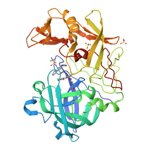
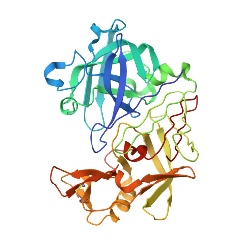
The Structure of Endothiapepsin Complexed with the Gem-Diol Inhibitor Pd-135,040 at 1.37 A

Coates, L.Erskine, P.T.Mall, S.Williams, P.A.Gill, R.S.Wood, S.P.Cooper, J.B.

(2003) Acta Crystallogr D Biol Crystallogr 59: 978

  • DOI: https://doi.org/10.1107/s0907444903006267
  • Primary Citation of Related Structures:  
    1OD1

  • PubMed Abstract: 

    The crystal structure of endothiapepsin complexed with the gem-diol inhibitor PD-135,040 has been anisotropically refined to a resolution of 1.37 A. The structure of this inhibitor complex is in agreement with previous structures of endothiapepsin gem-diol inhibitor complexes that have been used to develop proposed catalytic mechanisms. However, the increase in resolution over previous structures confirms the presence of a number of short hydrogen bonds within the active site that are likely to play an important role in the catalytic mechanism. The presence of low-barrier hydrogen bonds was indicated in a previous one-dimensional H NMR spectrum.


  • Organizational Affiliation

    School of Biological Sciences, University of Southampton, Bassett Crescent East, Southampton SO16 7PX, England. leightonc@bigfoot.com


Macromolecules
Find similar proteins by:  (by identity cutoff)  |  3D Structure
Entity ID: 1
MoleculeChains Sequence LengthOrganismDetailsImage
ENDOTHIAPEPSIN330Cryphonectria parasiticaMutation(s): 0 
EC: 3.4.23.22
UniProt
Find proteins for P11838 (Cryphonectria parasitica)
Explore P11838 
Go to UniProtKB:  P11838
Entity Groups  
Sequence Clusters30% Identity50% Identity70% Identity90% Identity95% Identity100% Identity
UniProt GroupP11838
Sequence Annotations
Expand
  • Reference Sequence
Small Molecules
Ligands 2 Unique
IDChains Name / Formula / InChI Key2D Diagram3D Interactions
0QS
Query on 0QS

Download Ideal Coordinates CCD File 
B [auth A]N~2~-[(2R)-2-benzyl-3-(tert-butylsulfonyl)propanoyl]-N-{(1R)-1-(cyclohexylmethyl)-3,3-difluoro-2,2-dihydroxy-4-[(2-morpholin-4-ylethyl)amino]-4-oxobutyl}-3-(1H-imidazol-3-ium-4-yl)-L-alaninamide
C36 H56 F2 N7 O8 S
RRJUDVVMLCYZDV-DTXPUJKBSA-O
SO4
Query on SO4

Download Ideal Coordinates CCD File 
C [auth A],
D [auth A]
SULFATE ION
O4 S
QAOWNCQODCNURD-UHFFFAOYSA-L
Biologically Interesting Molecules (External Reference) 1 Unique
Experimental Data & Validation

Experimental Data

  • Method: X-RAY DIFFRACTION
  • Resolution: 1.37 Å
  • R-Value Free:  0.159 (Depositor) 
  • R-Value Work:  0.133 (DCC) 
  • R-Value Observed: 0.125 (Depositor) 
Space Group: P 1 21 1
Unit Cell:
Length ( Å )Angle ( ˚ )
a = 53.201α = 90
b = 73.249β = 110.1
c = 45.993γ = 90
Software Package:
Software NamePurpose
SHELXL-97refinement
MOSFLMdata reduction
SCALAdata scaling

Structure Validation

View Full Validation Report


Ligand Structure Quality Assessment 

Created with Raphaël 2.3.0Worse 01 BetterLigand structure goodness of fit to experimental dataBest fitted 0QSClick on this verticalbar to view details

Entry History 

Deposition Data

Revision History  (Full details and data files)

  • Version 1.0: 2003-06-12
    Type: Initial release
  • Version 1.1: 2011-07-13
    Changes: Atomic model, Database references, Derived calculations, Non-polymer description, Structure summary, Version format compliance
  • Version 1.2: 2012-11-30
    Changes: Other
  • Version 1.3: 2024-10-23
    Changes: Data collection, Database references, Derived calculations, Other, Structure summary