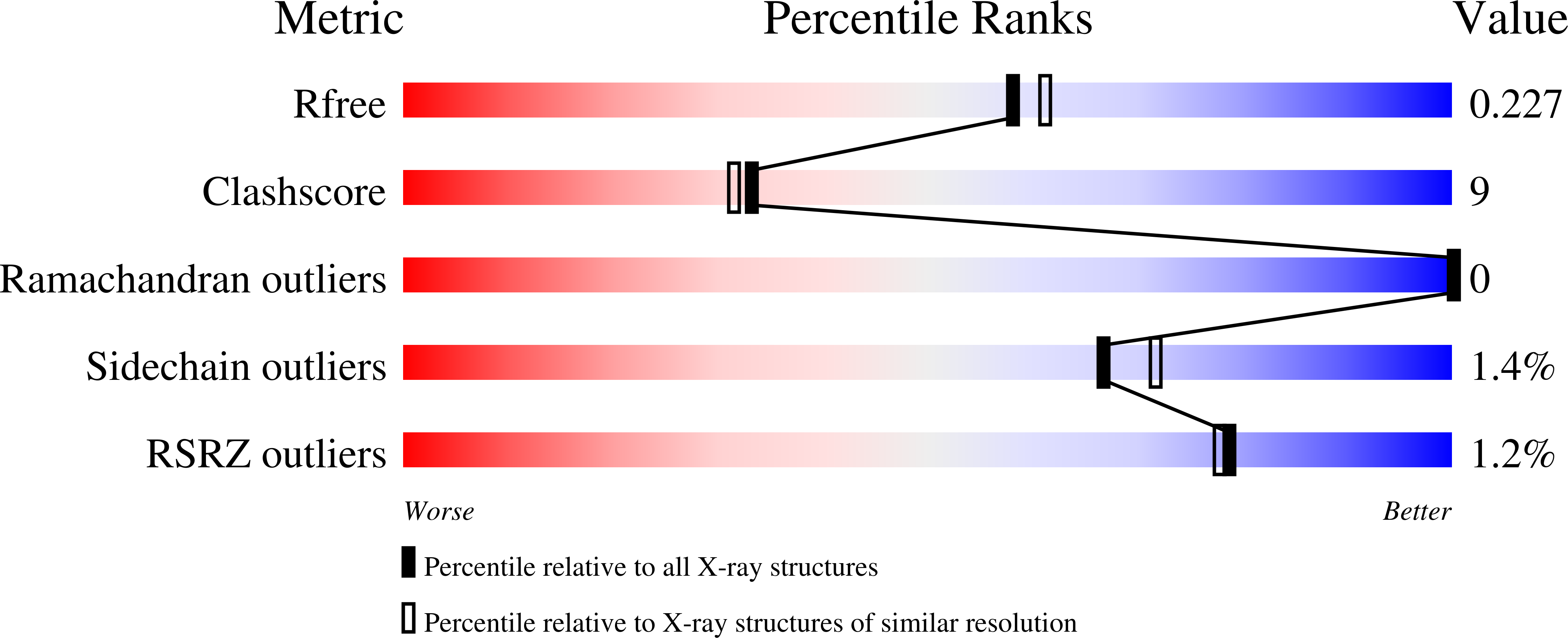
1HB6

Structure of bovine Acyl-CoA binding protein in orthorhombic crystal form


Experimental Data Snapshot

  • Method: X-RAY DIFFRACTION
  • Resolution: 2.00 Å
  • R-Value Free: 0.226 
  • R-Value Work: 0.200 
  • R-Value Observed: 0.200 

Starting Model: experimental
View more details

wwPDB Validation   3D Report Full Report


This is version 1.3 of the entry. See complete history


Literature

Binding Site Differences Revealed by Crystal Structures of Plasmodium Falciparum and Bovine Acyl-Coa Binding Protein

Van Aalten, D.M.F.Milne, K.G.Zou, J.Y.Kleywegt, G.J.Bergfors, T.Ferguson, M.A.J.Knudsen, J.Jones, T.A.

(2001) J Mol Biol 309: 181

  • DOI: https://doi.org/10.1006/jmbi.2001.4749
  • Primary Citation of Related Structures:  
    1HB6, 1HB8, 1HBK

  • PubMed Abstract: 

    Acyl-CoA binding protein (ACBP) maintains a pool of fatty acyl-CoA molecules in the cell and plays a role in fatty acid metabolism. The biochemical properties of Plasmodium falciparum ACBP are described together with the 2.0 A resolution crystal structures of a P. falciparum ACBP-acyl-CoA complex and of bovine ACBP in two crystal forms. Overall, the bovine ACBP crystal structures are similar to the NMR structures published previously; however, the bovine and parasite ACBP structures are less similar. The parasite ACBP is shown to have a different ligand-binding pocket, leading to an acyl-CoA binding specificity different from that of bovine ACBP. Several non-conservative differences in residues that interact with the ligand were identified between the mammalian and parasite ACBPs. These, together with measured binding-specificity differences, suggest that there is a potential for the design of molecules that might selectively block the acyl-CoA binding site.


  • Organizational Affiliation

    Wellcome Trust Biocentre, Department of Biochemistry University of Dundee, Scotland. dava@davapc1.bioch.dundee.ac.uk


Macromolecules
Find similar proteins by:  (by identity cutoff)  |  3D Structure
Entity ID: 1
MoleculeChains Sequence LengthOrganismDetailsImage
ACYL-COA BINDING PROTEIN86Bos taurusMutation(s): 0 
UniProt
Find proteins for P07107 (Bos taurus)
Explore P07107 
Go to UniProtKB:  P07107
Entity Groups  
Sequence Clusters30% Identity50% Identity70% Identity90% Identity95% Identity100% Identity
UniProt GroupP07107
Sequence Annotations
Expand
  • Reference Sequence
Small Molecules
Ligands 1 Unique
IDChains Name / Formula / InChI Key2D Diagram3D Interactions
CD
Query on CD

Download Ideal Coordinates CCD File 
B [auth A]CADMIUM ION
Cd
WLZRMCYVCSSEQC-UHFFFAOYSA-N
Experimental Data & Validation

Experimental Data

  • Method: X-RAY DIFFRACTION
  • Resolution: 2.00 Å
  • R-Value Free: 0.226 
  • R-Value Work: 0.200 
  • R-Value Observed: 0.200 
  • Space Group: P 21 21 21
Unit Cell:
Length ( Å )Angle ( ˚ )
a = 26.05α = 90
b = 54.78β = 90
c = 65.83γ = 90
Software Package:
Software NamePurpose
CNSrefinement
UCSDdata reduction
UCSDdata scaling
AMoREphasing

Structure Validation

View Full Validation Report


Entry History 

Deposition Data

Revision History  (Full details and data files)

  • Version 1.0: 2002-03-11
    Type: Initial release
  • Version 1.1: 2011-05-07
    Changes: Version format compliance
  • Version 1.2: 2011-07-13
    Changes: Version format compliance
  • Version 1.3: 2023-12-13
    Changes: Data collection, Database references, Derived calculations, Other, Refinement description