1UOD | pdb_00001uod

Crystal structure of the dihydroxyacetone kinase from E. coli in complex with dihydroxyacetone-phosphate


Experimental Data Snapshot

  • Method: X-RAY DIFFRACTION
  • Resolution: 1.90 Å
  • R-Value Free: 
    0.206 (Depositor) 
  • R-Value Work: 
    0.167 (Depositor), 0.189 (DCC) 
  • R-Value Observed: 
    0.168 (Depositor) 

wwPDB Validation   3D Report Full Report


Ligand Structure Quality Assessment 

Created with Raphaël 2.3.0Worse 01 BetterLigand structure goodness of fit to experimental dataBest fitted G3HClick on this verticalbar to view details

This is version 1.2 of the entry. See complete history


Literature

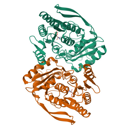
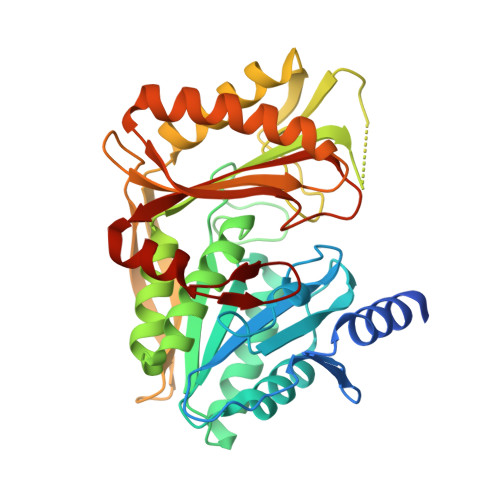
Phosphoenolpyruvate- and ATP-Dependent Dihydroxyacetone Kinases: Covalent Substrate-Binding and Kinetic Mechanism

Garcia-Alles, L.F.Siebold, C.Luthi-Nyffeler, T.Flukiger-Bruhwiler, K.Schneider, P.Burgi, H.-B.Baumann, U.Erni, B.

(2004) Biochemistry 43: 13037

  • DOI: https://doi.org/10.1021/bi048575m
  • Primary Citation of Related Structures:  
    1UOD, 1UOE

  • PubMed Abstract: 

    Dihydroxyacetone (Dha) kinases are a sequence-conserved family of enzymes, which utilize two different phosphoryldonors, ATP in animals, plants, and some bacteria, and a multiphosphoprotein of the phosphoenolpyruvate carbohydrate phosphotransferase system (PTS) in most bacteria. Here, we compare the PTS-dependent kinase of Escherichia coli and the ATP-dependent kinase of Citrobacter freundii. They display 30% sequence identity. The binding constants of the E. coli kinase for eleven short-chain carbonyl compounds were determined by acetone precipitation of the enzyme-substrate complexes. They are 3.4 microM for Dha, 780 microM for Dha-phosphate (DhaP), 50 microM for D,L-glyceraldehyde (GA), and 90 microM for D,L-glyceraldehyde-3-phosphate. The k(cat) for Dha of the PTS-dependent kinase is 290 min(-1), and that of the ATP-dependent kinase is 1050 min(-1). The Km for Dha of both kinases is <6 microM. The X-ray structures of the enzyme-GA and the enzyme-DhaP complex show that substrates as well as products are bound in hemiaminal linkage to an active-site histidine. Quantum-mechanical calculations offer no indication for activation of the reacting hydroxyl group by the formation of the hemiaminal. However, the formation of the hemiaminal bond allows selection for short-chain carbonyl compounds and discrimination against structurally similar polyols. The Dha kinase remains fully active in the presence of 2 M glycerol, and phosphorylates trace impurities of carbonyl compounds present in glycerol.


  • Organizational Affiliation

    Department of Chemistry and Biochemistry and Laboratory for Chemical and Mineralogical Crystallography, University of Berne, CH-3012 Berne, Switzerland.


Macromolecules
Find similar proteins by:  (by identity cutoff)  |  3D Structure
Entity ID: 1
MoleculeChains Sequence LengthOrganismDetailsImage
DIHYDROXYACETONE KINASE
A, B
366Escherichia coliMutation(s): 0 
EC: 2.7.1.29 (PDB Primary Data), 2.7.1.121 (UniProt)
UniProt
Find proteins for P76015 (Escherichia coli (strain K12))
Explore P76015 
Go to UniProtKB:  P76015
Entity Groups  
Sequence Clusters30% Identity50% Identity70% Identity90% Identity95% Identity100% Identity
UniProt GroupP76015
Sequence Annotations
Expand
  • Reference Sequence
Experimental Data & Validation

Experimental Data

  • Method: X-RAY DIFFRACTION
  • Resolution: 1.90 Å
  • R-Value Free:  0.206 (Depositor) 
  • R-Value Work:  0.167 (Depositor), 0.189 (DCC) 
  • R-Value Observed: 0.168 (Depositor) 
Space Group: P 21 21 2
Unit Cell:
Length ( Å )Angle ( ˚ )
a = 96.498α = 90
b = 97.055β = 90
c = 86.123γ = 90
Software Package:
Software NamePurpose
REFMACrefinement

Structure Validation

View Full Validation Report


Ligand Structure Quality Assessment 

Created with Raphaël 2.3.0Worse 01 BetterLigand structure goodness of fit to experimental dataBest fitted G3HClick on this verticalbar to view details

Entry History 

Deposition Data

Revision History  (Full details and data files)

  • Version 1.0: 2004-09-24
    Type: Initial release
  • Version 1.1: 2011-07-13
    Changes: Advisory, Version format compliance
  • Version 1.2: 2024-11-13
    Changes: Data collection, Database references, Derived calculations, Other, Refinement description, Structure summary