Phosphoenolpyruvate- and ATP-Dependent Dihydroxyacetone Kinases: Covalent Substrate-Binding and Kinetic Mechanism
Garcia-Alles, L.F., Siebold, C., Luthi-Nyffeler, T., Flukiger-Bruhwiler, K., Schneider, P., Burgi, H.-B., Baumann, U., Erni, B.(2004) Biochemistry 43: 13037
- PubMed: 15476397
- DOI: https://doi.org/10.1021/bi048575m
- Primary Citation of Related Structures:
1UOD, 1UOE - PubMed Abstract:
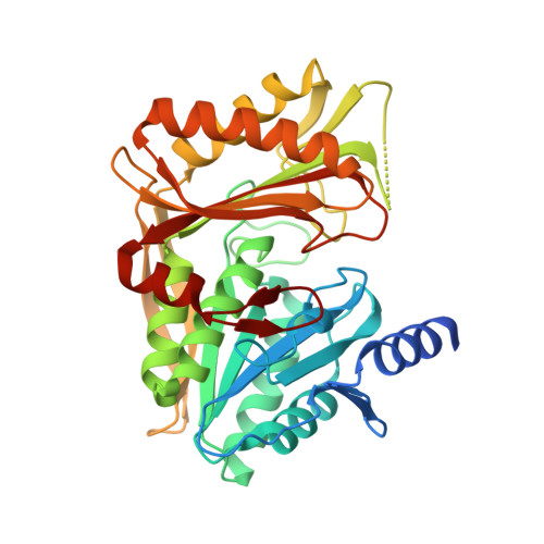
Dihydroxyacetone (Dha) kinases are a sequence-conserved family of enzymes, which utilize two different phosphoryldonors, ATP in animals, plants, and some bacteria, and a multiphosphoprotein of the phosphoenolpyruvate carbohydrate phosphotransferase system (PTS) in most bacteria. Here, we compare the PTS-dependent kinase of Escherichia coli and the ATP-dependent kinase of Citrobacter freundii. They display 30% sequence identity. The binding constants of the E. coli kinase for eleven short-chain carbonyl compounds were determined by acetone precipitation of the enzyme-substrate complexes. They are 3.4 microM for Dha, 780 microM for Dha-phosphate (DhaP), 50 microM for D,L-glyceraldehyde (GA), and 90 microM for D,L-glyceraldehyde-3-phosphate. The k(cat) for Dha of the PTS-dependent kinase is 290 min(-1), and that of the ATP-dependent kinase is 1050 min(-1). The Km for Dha of both kinases is <6 microM. The X-ray structures of the enzyme-GA and the enzyme-DhaP complex show that substrates as well as products are bound in hemiaminal linkage to an active-site histidine. Quantum-mechanical calculations offer no indication for activation of the reacting hydroxyl group by the formation of the hemiaminal. However, the formation of the hemiaminal bond allows selection for short-chain carbonyl compounds and discrimination against structurally similar polyols. The Dha kinase remains fully active in the presence of 2 M glycerol, and phosphorylates trace impurities of carbonyl compounds present in glycerol.
Organizational Affiliation:
Department of Chemistry and Biochemistry and Laboratory for Chemical and Mineralogical Crystallography, University of Berne, CH-3012 Berne, Switzerland.


