3SL8 | pdb_00003sl8

Crystal structure of the catalytic domain of PDE4D2 with compound 10o


Experimental Data Snapshot

  • Method: X-RAY DIFFRACTION
  • Resolution: 2.60 Å
  • R-Value Free: 
    0.273 (Depositor), 0.270 (DCC) 
  • R-Value Work: 
    0.196 (Depositor), 0.190 (DCC) 
  • R-Value Observed: 
    0.200 (Depositor) 

wwPDB Validation   3D Report Full Report


Ligand Structure Quality Assessment 

Created with Raphaël 2.3.0Worse 01 BetterLigand structure goodness of fit to experimental dataBest fitted JN7Click on this verticalbar to view details

This is version 1.3 of the entry. See complete history


Literature

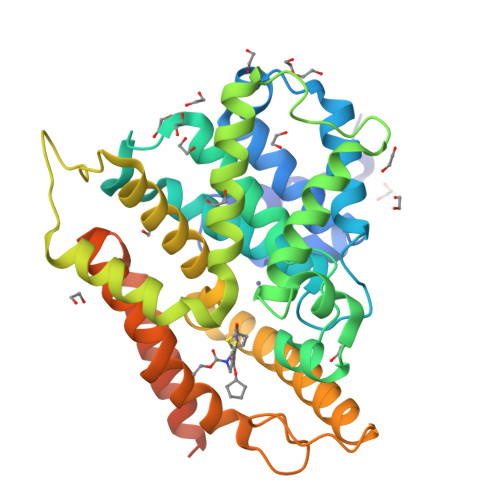
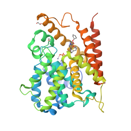
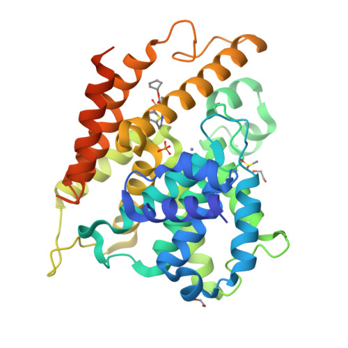
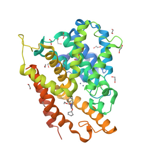
Thiophene inhibitors of PDE4: Crystal structures show a second binding mode at the catalytic domain of PDE4D2.

Nankervis, J.L.Feil, S.C.Hancock, N.C.Zheng, Z.Ng, H.L.Morton, C.J.Holien, J.K.Ho, P.W.Frazzetto, M.M.Jennings, I.G.Manallack, D.T.Martin, T.J.Thompson, P.E.Parker, M.W.

(2011) Bioorg Med Chem Lett 21: 7089-7093

  • DOI: https://doi.org/10.1016/j.bmcl.2011.09.109
  • Primary Citation of Related Structures:  
    3SL3, 3SL4, 3SL5, 3SL6, 3SL8

  • PubMed Abstract: 

    PDE4 inhibitors have been identified as therapeutic targets for a variety of conditions, particularly inflammatory diseases. We have serendipitously identified a novel class of phosphodiesterase 4 (PDE4) inhibitor during a study to discover antagonists of the parathyroid hormone receptor. X-ray crystallographic studies of PDE4D2 complexed to four potent inhibitors reveal the atomic details of how they inhibit the enzyme and a notable contrast to another recently reported thiophene-based inhibitor.


  • Organizational Affiliation

    Medicinal Chemistry & Drug Action, Monash Institute of Pharmaceutical Sciences, Monash University (Parkville campus), Parkville, Victoria, Australia.


Macromolecules
Find similar proteins by:  (by identity cutoff)  |  3D Structure
Entity ID: 1
MoleculeChains Sequence LengthOrganismDetailsImage
cAMP-specific 3',5'-cyclic phosphodiesterase 4D
A, B, C, D
361Homo sapiensMutation(s): 0 
Gene Names: PDE4DDPDE3
EC: 3.1.4.17 (PDB Primary Data), 3.1.4.53 (UniProt)
UniProt & NIH Common Fund Data Resources
Find proteins for Q08499 (Homo sapiens)
Explore Q08499 
Go to UniProtKB:  Q08499
PHAROS:  Q08499
GTEx:  ENSG00000113448 
Entity Groups  
Sequence Clusters30% Identity50% Identity70% Identity90% Identity95% Identity100% Identity
UniProt GroupQ08499
Sequence Annotations
Expand
  • Reference Sequence
Small Molecules
Ligands 6 Unique
IDChains Name / Formula / InChI Key2D Diagram3D Interactions
JN7
Query on JN7

Download Ideal Coordinates CCD File 
AA [auth B],
IA [auth C],
T [auth A],
ZA [auth D]
3-cyclopentyl 6-ethenyl 2-[(thiophen-2-ylacetyl)amino]-4,7-dihydrothieno[2,3-c]pyridine-3,6(5H)-dicarboxylate
C22 H24 N2 O5 S2
RIZICIJVRFSMJP-UHFFFAOYSA-N
PEG
Query on PEG

Download Ideal Coordinates CCD File 
N [auth A],
OA [auth D]
DI(HYDROXYETHYL)ETHER
C4 H10 O3
MTHSVFCYNBDYFN-UHFFFAOYSA-N
PO4
Query on PO4

Download Ideal Coordinates CCD File 
GA [auth C],
X [auth B]
PHOSPHATE ION
O4 P
NBIIXXVUZAFLBC-UHFFFAOYSA-K
DMS
Query on DMS

Download Ideal Coordinates CCD File 
HA [auth C],
XA [auth D]
DIMETHYL SULFOXIDE
C2 H6 O S
IAZDPXIOMUYVGZ-UHFFFAOYSA-N
ZN
Query on ZN

Download Ideal Coordinates CCD File 
EA [auth C]
F [auth A]
FA [auth C]
G [auth A]
PA [auth D]
ZINC ION
Zn
PTFCDOFLOPIGGS-UHFFFAOYSA-N
EDO
Query on EDO

Download Ideal Coordinates CCD File 
BA [auth C]
CA [auth C]
DA [auth C]
E [auth A]
H [auth A]
1,2-ETHANEDIOL
C2 H6 O2
LYCAIKOWRPUZTN-UHFFFAOYSA-N
Experimental Data & Validation

Experimental Data

  • Method: X-RAY DIFFRACTION
  • Resolution: 2.60 Å
  • R-Value Free:  0.273 (Depositor), 0.270 (DCC) 
  • R-Value Work:  0.196 (Depositor), 0.190 (DCC) 
  • R-Value Observed: 0.200 (Depositor) 
Space Group: P 21 21 21
Unit Cell:
Length ( Å )Angle ( ˚ )
a = 99.14α = 90
b = 111.65β = 90
c = 161.74γ = 90
Software Package:
Software NamePurpose
XSCALEdata scaling
PHENIXrefinement
PDB_EXTRACTdata extraction
Blu-Icedata collection
XDSdata reduction
XDSdata scaling
MOLREPphasing

Structure Validation

View Full Validation Report


Ligand Structure Quality Assessment 

Created with Raphaël 2.3.0Worse 01 BetterLigand structure goodness of fit to experimental dataBest fitted JN7Click on this verticalbar to view details

Entry History 

Deposition Data

  • Released Date: 2011-10-26 
  • Deposition Author(s): Feil, S.F.

Revision History  (Full details and data files)

  • Version 1.0: 2011-10-26
    Type: Initial release
  • Version 1.1: 2011-12-14
    Changes: Database references
  • Version 1.2: 2017-11-08
    Changes: Refinement description
  • Version 1.3: 2024-02-28
    Changes: Data collection, Database references, Derived calculations