4C2Y | pdb_00004c2y

Human N-myristoyltransferase (NMT1) with Myristoyl-CoA co-factor


Experimental Data Snapshot

  • Method: X-RAY DIFFRACTION
  • Resolution: 1.64 Å
  • R-Value Free: 
    0.222 (Depositor), 0.220 (DCC) 
  • R-Value Work: 
    0.170 (Depositor), 0.170 (DCC) 
  • R-Value Observed: 
    0.173 (Depositor) 

Starting Model: experimental
View more details

wwPDB Validation   3D Report Full Report


Ligand Structure Quality Assessment 

Created with Raphaël 2.3.0Worse 01 BetterLigand structure goodness of fit to experimental dataBest fitted MYAClick on this verticalbar to view details

This is version 1.2 of the entry. See complete history


Literature

Global Profiling of Co- and Post-Translationally N-Myristoylated Proteomes in Human Cells.

Thinon, E.Serwa, R.A.Broncel, M.Brannigan, J.A.Brassat, U.Wright, M.H.Heal, W.P.Wilkinson, A.J.Mann, D.J.Tate, E.W.

(2014) Nat Commun 5: 4919

  • DOI: https://doi.org/10.1038/ncomms5919
  • Primary Citation of Related Structures:  
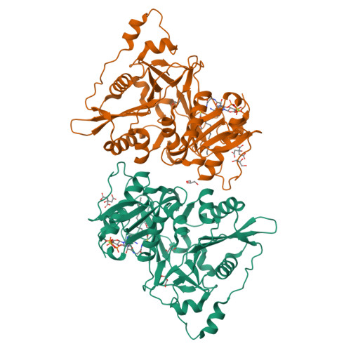
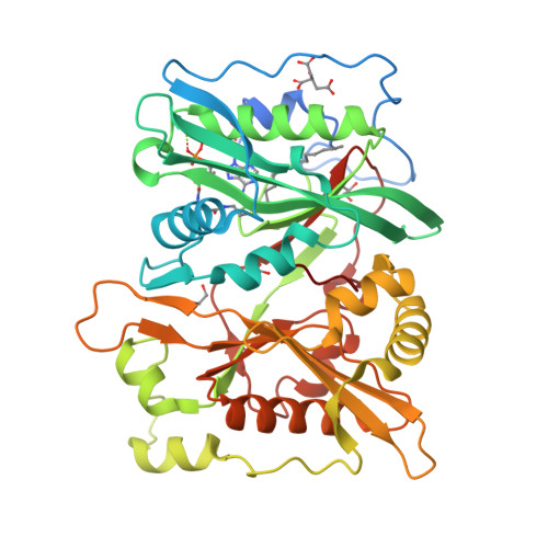
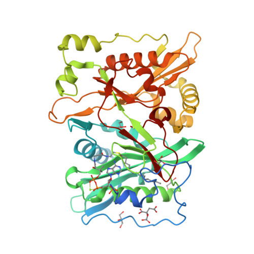
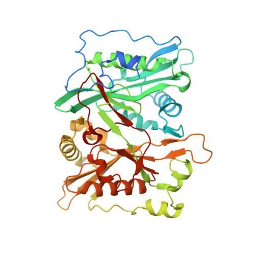
    4C2X, 4C2Y, 4C2Z

  • PubMed Abstract: 

    Protein N-myristoylation is a ubiquitous co- and post-translational modification that has been implicated in the development and progression of a range of human diseases. Here, we report the global N-myristoylated proteome in human cells determined using quantitative chemical proteomics combined with potent and specific human N-myristoyltransferase (NMT) inhibition. Global quantification of N-myristoylation during normal growth or apoptosis allowed the identification of >100 N-myristoylated proteins, >95% of which are identified for the first time at endogenous levels. Furthermore, quantitative dose response for inhibition of N-myristoylation is determined for >70 substrates simultaneously across the proteome. Small-molecule inhibition through a conserved substrate-binding pocket is also demonstrated by solving the crystal structures of inhibitor-bound NMT1 and NMT2. The presented data substantially expand the known repertoire of co- and post-translational N-myristoylation in addition to validating tools for the pharmacological inhibition of NMT in living cells.


  • Organizational Affiliation

    1] Department of Chemistry, Imperial College London, Exhibition Road, London SW7 2AZ, UK [2] Department of Life Sciences, Imperial College London, Exhibition Road, London SW7 2AZ, UK.


Macromolecules
Find similar proteins by:  (by identity cutoff)  |  3D Structure
Entity ID: 1
MoleculeChains Sequence LengthOrganismDetailsImage
GLYCYLPEPTIDE N-TETRADECANOYLTRANSFERASE 1
A, B
410Homo sapiensMutation(s): 0 
EC: 2.3.1.97 (PDB Primary Data), 2.3.1 (UniProt)
UniProt & NIH Common Fund Data Resources
Find proteins for P30419 (Homo sapiens)
Explore P30419 
Go to UniProtKB:  P30419
PHAROS:  P30419
GTEx:  ENSG00000136448 
Entity Groups  
Sequence Clusters30% Identity50% Identity70% Identity90% Identity95% Identity100% Identity
UniProt GroupP30419
Sequence Annotations
Expand
  • Reference Sequence
Small Molecules
Ligands 4 Unique
IDChains Name / Formula / InChI Key2D Diagram3D Interactions
MYA
Query on MYA

Download Ideal Coordinates CCD File 
C [auth A],
I [auth B]
TETRADECANOYL-COA
C35 H62 N7 O17 P3 S
DUAFKXOFBZQTQE-QSGBVPJFSA-N
CIT
Query on CIT

Download Ideal Coordinates CCD File 
E [auth A],
K [auth B]
CITRIC ACID
C6 H8 O7
KRKNYBCHXYNGOX-UHFFFAOYSA-N
GOL
Query on GOL

Download Ideal Coordinates CCD File 
F [auth A]
G [auth A]
H [auth A]
L [auth B]
M [auth B]
GLYCEROL
C3 H8 O3
PEDCQBHIVMGVHV-UHFFFAOYSA-N
MG
Query on MG

Download Ideal Coordinates CCD File 
D [auth A],
J [auth B]
MAGNESIUM ION
Mg
JLVVSXFLKOJNIY-UHFFFAOYSA-N
Experimental Data & Validation

Experimental Data

  • Method: X-RAY DIFFRACTION
  • Resolution: 1.64 Å
  • R-Value Free:  0.222 (Depositor), 0.220 (DCC) 
  • R-Value Work:  0.170 (Depositor), 0.170 (DCC) 
  • R-Value Observed: 0.173 (Depositor) 
Space Group: P 21 21 2
Unit Cell:
Length ( Å )Angle ( ˚ )
a = 78.74α = 90
b = 178.48β = 90
c = 58.55γ = 90
Software Package:
Software NamePurpose
REFMACrefinement
XDSdata reduction
SCALAdata scaling
MOLREPphasing

Structure Validation

View Full Validation Report


Ligand Structure Quality Assessment 

Created with Raphaël 2.3.0Worse 01 BetterLigand structure goodness of fit to experimental dataBest fitted MYAClick on this verticalbar to view details

Entry History 

Deposition Data

Revision History  (Full details and data files)

  • Version 1.0: 2014-10-01
    Type: Initial release
  • Version 1.1: 2014-10-08
    Changes: Database references
  • Version 1.2: 2023-12-20
    Changes: Data collection, Database references, Derived calculations, Other, Refinement description