4C2Z

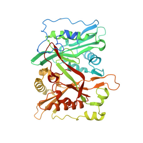
Human N-myristoyltransferase (NMT1) with Myristoyl-CoA and inhibitor bound


Experimental Data Snapshot

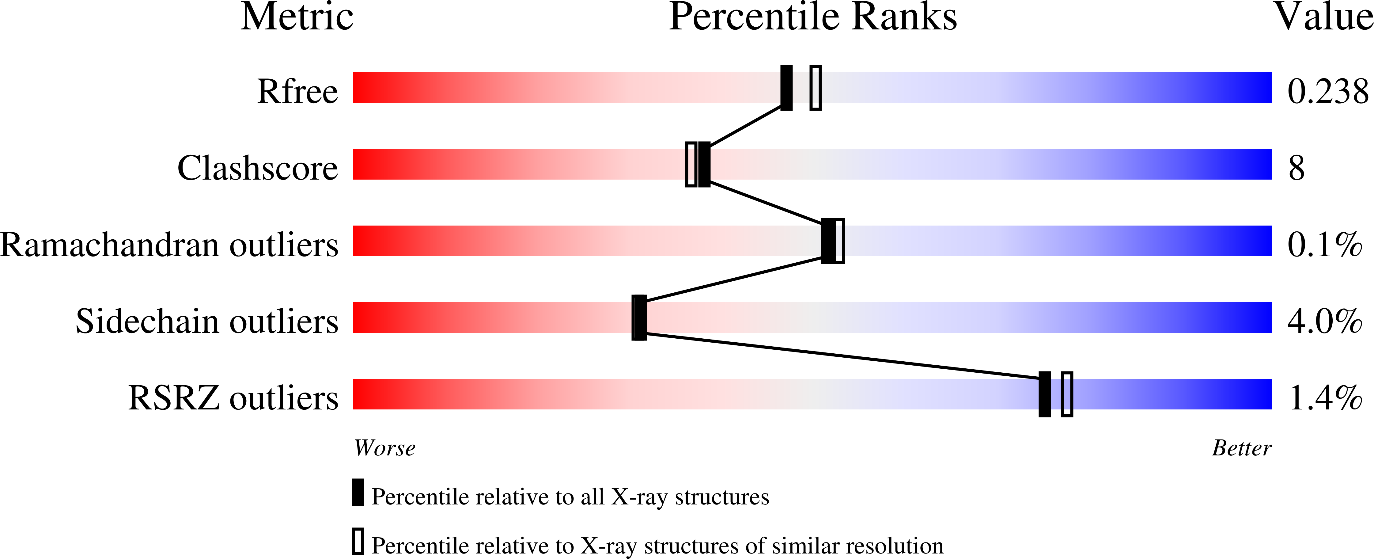
  • Method: X-RAY DIFFRACTION
  • Resolution: 2.08 Å
  • R-Value Free: 0.238 
  • R-Value Work: 0.169 
  • R-Value Observed: 0.173 

wwPDB Validation   3D Report Full Report


Ligand Structure Quality Assessment 


This is version 1.2 of the entry. See complete history


Literature

Chemical Proteomics Defines the Mammalian N- Myristoylated Proteome in Live Cells Global Profiling of Co- and Post-Translationally N-Myristoylated Proteomes in Human Cells.

Thinon, E.Serwa, R.A.Broncel, M.Brannigan, J.A.Brassat, U.Wright, M.H.Heal, W.P.Wilkinson, A.J.Mann, D.J.Tate, E.W.

(2014) Nat Commun 5: 4919

  • DOI: https://doi.org/10.1038/ncomms5919
  • Primary Citation of Related Structures:  
    4C2X, 4C2Y, 4C2Z

  • PubMed Abstract: 

    Protein N-myristoylation is a ubiquitous co- and post-translational modification that has been implicated in the development and progression of a range of human diseases. Here, we report the global N-myristoylated proteome in human cells determined using quantitative chemical proteomics combined with potent and specific human N-myristoyltransferase (NMT) inhibition. Global quantification of N-myristoylation during normal growth or apoptosis allowed the identification of >100 N-myristoylated proteins, >95% of which are identified for the first time at endogenous levels. Furthermore, quantitative dose response for inhibition of N-myristoylation is determined for >70 substrates simultaneously across the proteome. Small-molecule inhibition through a conserved substrate-binding pocket is also demonstrated by solving the crystal structures of inhibitor-bound NMT1 and NMT2. The presented data substantially expand the known repertoire of co- and post-translational N-myristoylation in addition to validating tools for the pharmacological inhibition of NMT in living cells.


  • Organizational Affiliation

    1] Department of Chemistry, Imperial College London, Exhibition Road, London SW7 2AZ, UK [2] Department of Life Sciences, Imperial College London, Exhibition Road, London SW7 2AZ, UK.


Macromolecules
Find similar proteins by:  (by identity cutoff)  |  3D Structure
Entity ID: 1
MoleculeChains Sequence LengthOrganismDetailsImage
GLYCYLPEPTIDE N-TETRADECANOYLTRANSFERASE 1
A, B
410Homo sapiensMutation(s): 0 
EC: 2.3.1.97 (PDB Primary Data), 2.3.1 (UniProt)
UniProt & NIH Common Fund Data Resources
Find proteins for P30419 (Homo sapiens)
Explore P30419 
Go to UniProtKB:  P30419
PHAROS:  P30419
GTEx:  ENSG00000136448 
Entity Groups  
Sequence Clusters30% Identity50% Identity70% Identity90% Identity95% Identity100% Identity
UniProt GroupP30419
Sequence Annotations
Expand
  • Reference Sequence
Small Molecules
Ligands 6 Unique
IDChains Name / Formula / InChI Key2D Diagram3D Interactions
MYA
Query on MYA

Download Ideal Coordinates CCD File 
C [auth A],
L [auth B]
TETRADECANOYL-COA
C35 H62 N7 O17 P3 S
DUAFKXOFBZQTQE-QSGBVPJFSA-N
646
Query on 646

Download Ideal Coordinates CCD File 
D [auth A],
M [auth B]
2,6-dichloro-4-(2-piperazin-1-ylpyridin-4-yl)-N-(1,3,5-trimethyl-1H-pyrazol-4-yl)benzenesulfonamide
C21 H24 Cl2 N6 O2 S
XMBSZPZJLPTFMV-UHFFFAOYSA-N
CIT
Query on CIT

Download Ideal Coordinates CCD File 
F [auth A],
O [auth B]
CITRIC ACID
C6 H8 O7
KRKNYBCHXYNGOX-UHFFFAOYSA-N
GOL
Query on GOL

Download Ideal Coordinates CCD File 
G [auth A],
H [auth A],
P [auth B],
Q [auth B]
GLYCEROL
C3 H8 O3
PEDCQBHIVMGVHV-UHFFFAOYSA-N
CL
Query on CL

Download Ideal Coordinates CCD File 
I [auth A]
J [auth A]
K [auth A]
R [auth B]
S [auth B]
I [auth A],
J [auth A],
K [auth A],
R [auth B],
S [auth B],
T [auth B],
U [auth B],
V [auth B]
CHLORIDE ION
Cl
VEXZGXHMUGYJMC-UHFFFAOYSA-M
MG
Query on MG

Download Ideal Coordinates CCD File 
E [auth A],
N [auth B]
MAGNESIUM ION
Mg
JLVVSXFLKOJNIY-UHFFFAOYSA-N
Experimental Data & Validation

Experimental Data

  • Method: X-RAY DIFFRACTION
  • Resolution: 2.08 Å
  • R-Value Free: 0.238 
  • R-Value Work: 0.169 
  • R-Value Observed: 0.173 
  • Space Group: P 21 21 2
Unit Cell:
Length ( Å )Angle ( ˚ )
a = 78.63α = 90
b = 179.12β = 90
c = 58.59γ = 90
Software Package:
Software NamePurpose
REFMACrefinement
XDSdata reduction
SCALAdata scaling

Structure Validation

View Full Validation Report


Ligand Structure Quality Assessment 


Entry History 

Deposition Data

Revision History  (Full details and data files)

  • Version 1.0: 2014-10-01
    Type: Initial release
  • Version 1.1: 2014-10-08
    Changes: Database references
  • Version 1.2: 2024-05-08
    Changes: Data collection, Database references, Derived calculations, Other