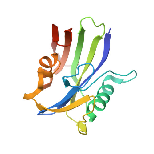
MTH1 Substrate Recognition--An Example of Specific Promiscuity.
Nissink, J.W., Bista, M., Breed, J., Carter, N., Embrey, K., Read, J., Winter-Holt, J.J.(2016) PLoS One 11: e0151154-e0151154
- PubMed: 26999531
- DOI: https://doi.org/10.1371/journal.pone.0151154
- Primary Citation of Related Structures:
5FSI, 5FSK, 5FSL, 5FSM, 5FSN, 5FSO - PubMed Abstract:
MTH1 (NUDT1) is an oncologic target involved in the prevention of DNA damage. We investigate the way MTH1 recognises its substrates and present substrate-bound structures of MTH1 for 8-oxo-dGTP and 8-oxo-rATP as examples of novel strong and weak binding substrate motifs. Investigation of a small set of purine-like fragments using 2D NMR resulted in identification of a fragment with weak potency. The protein-ligand X-Ray structure of this fragment provides insight into the role of water molecules in substrate selectivity. Wider fragment screening by NMR resulted in three new protein structures exhibiting alternative binding configurations to the key Asp-Asp recognition element of the protein. These inhibitor binding modes demonstrate that MTH1 employs an intricate yet promiscuous mechanism of substrate anchoring through its Asp-Asp pharmacophore. The structures suggest that water-mediated interactions convey selectivity towards oxidized substrates over their non-oxidised counterparts, in particular by stabilization of a water molecule in a hydrophobic environment through hydrogen bonding. These findings may be useful in the design of inhibitors of MTH1.
Organizational Affiliation:
Chemistry, Oncology, Innovative Medicines and Early Development Biotech Unit, AstraZeneca, Unit 310 (Darwin Building), Cambridge Science Park, Milton Road, Cambridge, CB4 0WG, United Kingdom, and Alderley Park, Cheshire, SK10 4TG, United Kingdom.


