7BQ2

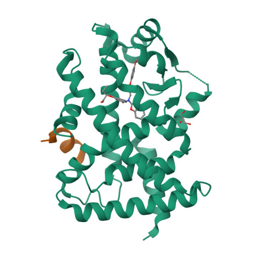
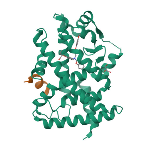
X-ray structure of human PPARalpha ligand binding domain-pemafibrate-SRC1 coactivator peptide co-crystals obtained by soaking


Experimental Data Snapshot

  • Method: X-RAY DIFFRACTION
  • Resolution: 1.52 Å
  • R-Value Free: 0.206 
  • R-Value Work: 0.182 
  • R-Value Observed: 0.184 

Starting Model: experimental
View more details

wwPDB Validation   3D Report Full Report


Ligand Structure Quality Assessment 


This is version 1.3 of the entry. See complete history


Literature

PPAR alpha Ligand-Binding Domain Structures with Endogenous Fatty Acids and Fibrates.

Kamata, S.Oyama, T.Saito, K.Honda, A.Yamamoto, Y.Suda, K.Ishikawa, R.Itoh, T.Watanabe, Y.Shibata, T.Uchida, K.Suematsu, M.Ishii, I.

(2020) iScience 23: 101727-101727

  • DOI: https://doi.org/10.1016/j.isci.2020.101727
  • Primary Citation of Related Structures:  
    6KAX, 6KAY, 6KAZ, 6KB0, 6KB1, 6KB2, 6KB3, 6KB4, 6KB5, 6KB6, 6KB7, 6KB8, 6KB9, 6KBA, 6KYP, 6L36, 6L37, 6L38, 6LX4, 6LX5, 6LX6, 6LX7, 6LX8, 6LX9, 6LXA, 6LXB, 6LXC, 7BPY, 7BPZ, 7BQ0, 7BQ1, 7BQ2, 7BQ3, 7BQ4

  • PubMed Abstract: 

    Most triacylglycerol-lowering fibrates have been developed in the 1960s-1980s before their molecular target, peroxisome proliferator-activated receptor alpha (PPARα), was identified. Twenty-one ligand-bound PPARα structures have been deposited in the Protein Data Bank since 2001; however, binding modes of fibrates and physiological ligands remain unknown. Here we show thirty-four X-ray crystallographic structures of the PPARα ligand-binding domain, which are composed of a "Center" and four "Arm" regions, in complexes with five endogenous fatty acids, six fibrates in clinical use, and six synthetic PPARα agonists. High-resolution structural analyses, in combination with coactivator recruitment and thermostability assays, demonstrate that stearic and palmitic acids are presumably physiological ligands; coordination to Arm III is important for high PPARα potency/selectivity of pemafibrate and GW7647; and agonistic activities of four fibrates are enhanced by the partial agonist GW9662. These results renew our understanding of PPARα ligand recognition and contribute to the molecular design of next-generation PPAR-targeted drugs.


  • Organizational Affiliation

    Laboratory of Health Chemistry, Showa Pharmaceutical University, Machida, Tokyo 194-8543, Japan.


Macromolecules
Find similar proteins by:  (by identity cutoff)  |  3D Structure
Entity ID: 1
MoleculeChains Sequence LengthOrganismDetailsImage
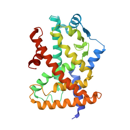
Peroxisome proliferator-activated receptor alpha273Homo sapiensMutation(s): 0 
UniProt & NIH Common Fund Data Resources
Find proteins for Q07869 (Homo sapiens)
Go to UniProtKB:  Q07869
PHAROS:  Q07869
GTEx:  ENSG00000186951 
Sequence Annotations
Expand
  • Reference Sequence

Find similar proteins by:  Sequence   |   3D Structure  

Entity ID: 2
MoleculeChains Sequence LengthOrganismDetailsImage
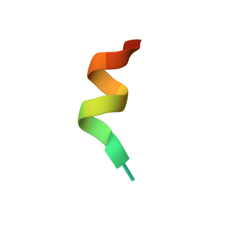
15-meric peptide from Nuclear receptor coactivator 115Homo sapiensMutation(s): 0 
EC: 2.3.1.48
UniProt & NIH Common Fund Data Resources
Find proteins for Q15788 (Homo sapiens)
Go to UniProtKB:  Q15788
PHAROS:  Q15788
GTEx:  ENSG00000084676 
Sequence Annotations
Expand
  • Reference Sequence
Small Molecules
Ligands 2 Unique
IDChains Name / Formula / InChI Key2D Diagram3D Interactions
P7F (Subject of Investigation/LOI)
Query on P7F

Download Ideal Coordinates CCD File 
C [auth A](2~{R})-2-[3-[[1,3-benzoxazol-2-yl-[3-(4-methoxyphenoxy)propyl]amino]methyl]phenoxy]butanoic acid
C28 H30 N2 O6
ZHKNLJLMDFQVHJ-RUZDIDTESA-N
GOL
Query on GOL

Download Ideal Coordinates CCD File 
D [auth A]GLYCEROL
C3 H8 O3
PEDCQBHIVMGVHV-UHFFFAOYSA-N
Binding Affinity Annotations 
IDSourceBinding Affinity
P7F BindingDB:  7BQ2 EC50: 1 (nM) from 1 assay(s)
Experimental Data & Validation

Experimental Data

  • Method: X-RAY DIFFRACTION
  • Resolution: 1.52 Å
  • R-Value Free: 0.206 
  • R-Value Work: 0.182 
  • R-Value Observed: 0.184 
  • Space Group: P 1 21 1
Unit Cell:
Length ( Å )Angle ( ˚ )
a = 44.977α = 90
b = 61.107β = 107.07
c = 53.206γ = 90
Software Package:
Software NamePurpose
XDSdata reduction
Aimlessdata scaling
PHASERphasing
PHENIXrefinement
PDB_EXTRACTdata extraction

Structure Validation

View Full Validation Report


Ligand Structure Quality Assessment 


Entry History & Funding Information

Deposition Data


Funding OrganizationLocationGrant Number
Ministry of Education, Culture, Sports, Science and Technology (Japan)Japan16H05107

Revision History  (Full details and data files)

  • Version 1.0: 2020-11-11
    Type: Initial release
  • Version 1.1: 2020-11-25
    Changes: Structure summary
  • Version 1.2: 2020-12-02
    Changes: Database references
  • Version 1.3: 2023-11-29
    Changes: Data collection, Database references, Refinement description