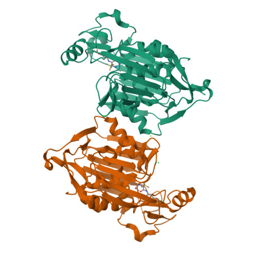
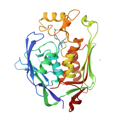
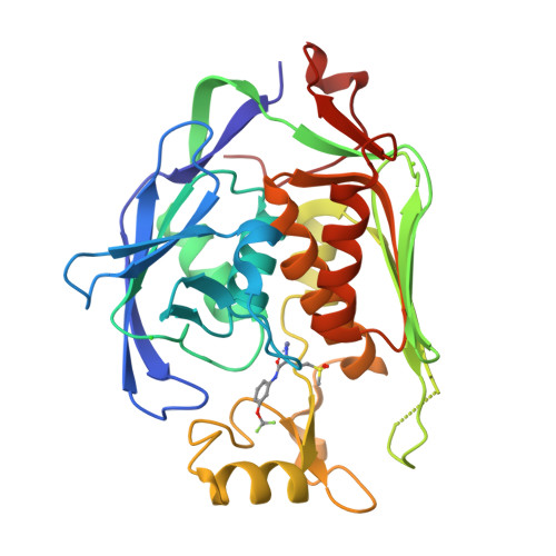
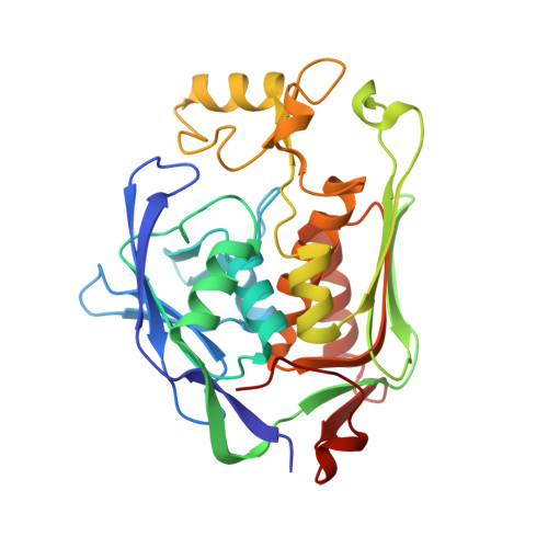
7CI4

Crystal structure of P.aeruginosa LpxC in complex with inhibitor


Experimental Data Snapshot

  • Method: X-RAY DIFFRACTION
  • Resolution: 2.00 Å
  • R-Value Free: 0.241 
  • R-Value Work: 0.187 
  • R-Value Observed: 0.190 

Starting Model: experimental
View more details

wwPDB Validation   3D Report Full Report


Ligand Structure Quality Assessment 


This is version 1.2 of the entry. See complete history


Literature

Fragment-Based Discovery of Novel Non-Hydroxamate LpxC Inhibitors with Antibacterial Activity.

Yamada, Y.Takashima, H.Walmsley, D.L.Ushiyama, F.Matsuda, Y.Kanazawa, H.Yamaguchi-Sasaki, T.Tanaka-Yamamoto, N.Yamagishi, J.Kurimoto-Tsuruta, R.Ogata, Y.Ohtake, N.Angove, H.Baker, L.Harris, R.Macias, A.Robertson, A.Surgenor, A.Watanabe, H.Nakano, K.Mima, M.Iwamoto, K.Okada, A.Takata, I.Hitaka, K.Tanaka, A.Fujita, K.Sugiyama, H.Hubbard, R.E.

(2020) J Med Chem 63: 14805-14820

  • DOI: https://doi.org/10.1021/acs.jmedchem.0c01215
  • Primary Citation of Related Structures:  
    7CI4, 7CI5, 7CI6, 7CI7, 7CI8, 7CI9, 7CIA, 7CIB, 7CIC, 7CID, 7CIE

  • PubMed Abstract: 

    UDP-3- O -acyl- N -acetylglucosamine deacetylase (LpxC) is a zinc metalloenzyme that catalyzes the first committed step in the biosynthesis of Lipid A, an essential component of the cell envelope of Gram-negative bacteria. The most advanced, disclosed LpxC inhibitors showing antibacterial activity coordinate zinc through a hydroxamate moiety with concerns about binding to other metalloenzymes. Here, we describe the discovery, optimization, and efficacy of two series of compounds derived from fragments with differing modes of zinc chelation. A series was evolved from a fragment where a glycine moiety complexes zinc, which achieved low nanomolar potency in an enzyme functional assay but poor antibacterial activity on cell cultures. A second series was based on a fragment that chelated zinc through an imidazole moiety. Structure-guided design led to a 2-(1 S -hydroxyethyl)-imidazole derivative exhibiting low nanomolar inhibition of LpxC and a minimum inhibitory concentration (MIC) of 4 μg/mL against Pseudomonas aeruginosa , which is little affected by the presence of albumin.


  • Organizational Affiliation

    Taisho Pharmaceutical Co., Ltd., Saitama 331-9530, Japan.


Macromolecules
Find similar proteins by:  (by identity cutoff)  |  3D Structure
Entity ID: 1
MoleculeChains Sequence LengthOrganismDetailsImage
UDP-3-O-acyl-N-acetylglucosamine deacetylase
A, B
299Pseudomonas aeruginosa PAO1Mutation(s): 1 
Gene Names: lpxCenvAPA4406
EC: 3.5.1.108
UniProt
Find proteins for P47205 (Pseudomonas aeruginosa (strain ATCC 15692 / DSM 22644 / CIP 104116 / JCM 14847 / LMG 12228 / 1C / PRS 101 / PAO1))
Explore P47205 
Go to UniProtKB:  P47205
Entity Groups  
Sequence Clusters30% Identity50% Identity70% Identity90% Identity95% Identity100% Identity
UniProt GroupP47205
Sequence Annotations
Expand
  • Reference Sequence
Small Molecules
Ligands 4 Unique
IDChains Name / Formula / InChI Key2D Diagram3D Interactions
FXU (Subject of Investigation/LOI)
Query on FXU

Download Ideal Coordinates CCD File 
C [auth A],
G [auth B]
(2R)-2-azanyl-4-methylsulfonyl-N-[3-(trifluoromethyloxy)phenyl]butanamide
C12 H15 F3 N2 O4 S
SJCAGVRRCBPLMJ-SNVBAGLBSA-N
ZN
Query on ZN

Download Ideal Coordinates CCD File 
D [auth A],
H [auth B]
ZINC ION
Zn
PTFCDOFLOPIGGS-UHFFFAOYSA-N
CL
Query on CL

Download Ideal Coordinates CCD File 
F [auth A],
J [auth B]
CHLORIDE ION
Cl
VEXZGXHMUGYJMC-UHFFFAOYSA-M
MG
Query on MG

Download Ideal Coordinates CCD File 
E [auth A],
I [auth B]
MAGNESIUM ION
Mg
JLVVSXFLKOJNIY-UHFFFAOYSA-N
Experimental Data & Validation

Experimental Data

  • Method: X-RAY DIFFRACTION
  • Resolution: 2.00 Å
  • R-Value Free: 0.241 
  • R-Value Work: 0.187 
  • R-Value Observed: 0.190 
  • Space Group: P 1 21 1
Unit Cell:
Length ( Å )Angle ( ˚ )
a = 68.367α = 90
b = 51.18β = 108.22
c = 96.763γ = 90
Software Package:
Software NamePurpose
REFMACrefinement
CrystalCleardata reduction
CrystalCleardata scaling
MOLREPphasing

Structure Validation

View Full Validation Report


Ligand Structure Quality Assessment 


Entry History 

Deposition Data

Revision History  (Full details and data files)

  • Version 1.0: 2020-12-02
    Type: Initial release
  • Version 1.1: 2020-12-23
    Changes: Database references
  • Version 1.2: 2023-11-29
    Changes: Data collection, Database references, Refinement description