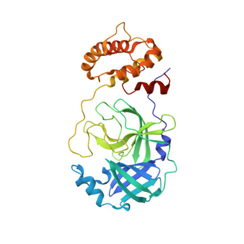
7GS6

Crystal structure of SARS-CoV-2 main protease in complex with cpd-29


Experimental Data Snapshot

  • Method: X-RAY DIFFRACTION
  • Resolution: 1.62 Å
  • R-Value Free: 0.228 
  • R-Value Work: 0.190 
  • R-Value Observed: 0.192 

Starting Model: experimental
View more details

wwPDB Validation   3D Report Full Report


Ligand Structure Quality Assessment 


This is version 1.2 of the entry. See complete history


Literature

Fragment-based screening targeting an open form of the SARS-CoV-2 main protease binding pocket.

Huang, C.Y.Metz, A.Lange, R.Artico, N.Potot, C.Hazemann, J.Muller, M.Dos Santos, M.Chambovey, A.Ritz, D.Eris, D.Meyer, S.Bourquin, G.Sharpe, M.Mac Sweeney, A.

(2024) Acta Crystallogr D Struct Biol 80: 123-136

  • DOI: https://doi.org/10.1107/S2059798324000329
  • Primary Citation of Related Structures:  
    7GRE, 7GRF, 7GRG, 7GRH, 7GRI, 7GRJ, 7GRK, 7GRL, 7GRM, 7GRN, 7GRO, 7GRP, 7GRQ, 7GRR, 7GRS, 7GRT, 7GRU, 7GRV, 7GRW, 7GRX, 7GRY, 7GRZ, 7GS0, 7GS1, 7GS2, 7GS3, 7GS4, 7GS5, 7GS6

  • PubMed Abstract: 

    To identify starting points for therapeutics targeting SARS-CoV-2, the Paul Scherrer Institute and Idorsia decided to collaboratively perform an X-ray crystallographic fragment screen against its main protease. Fragment-based screening was carried out using crystals with a pronounced open conformation of the substrate-binding pocket. Of 631 soaked fragments, a total of 29 hits bound either in the active site (24 hits), a remote binding pocket (three hits) or at crystal-packing interfaces (two hits). Notably, two fragments with a pose that was sterically incompatible with a more occluded crystal form were identified. Two isatin-based electrophilic fragments bound covalently to the catalytic cysteine residue. The structures also revealed a surprisingly strong influence of the crystal form on the binding pose of three published fragments used as positive controls, with implications for fragment screening by crystallography.


  • Organizational Affiliation

    Swiss Light Source, Paul Scherrer Institute, 5232 Villigen PSI, Switzerland.


Macromolecules
Find similar proteins by:  (by identity cutoff)  |  3D Structure
Entity ID: 1
MoleculeChains Sequence LengthOrganismDetailsImage
3C-like proteinase nsp5
A, B
306Severe acute respiratory syndrome coronavirus 2Mutation(s): 0 
Gene Names: rep1a-1b
EC: 3.4.22.69
UniProt
Find proteins for P0DTD1 (Severe acute respiratory syndrome coronavirus 2)
Explore P0DTD1 
Go to UniProtKB:  P0DTD1
Entity Groups  
Sequence Clusters30% Identity50% Identity70% Identity90% Identity95% Identity100% Identity
UniProt GroupP0DTD1
Sequence Annotations
Expand
  • Reference Sequence
Small Molecules
Ligands 4 Unique
IDChains Name / Formula / InChI Key2D Diagram3D Interactions
XXN (Subject of Investigation/LOI)
Query on XXN

Download Ideal Coordinates CCD File 
D [auth A],
I [auth B],
J [auth B]
(3S)-4,7-dichloro-3-hydroxy-1,3-dihydro-2H-indol-2-one
C8 H5 Cl2 N O2
JSUBYQLEJVQEIU-ZETCQYMHSA-N
SO4
Query on SO4

Download Ideal Coordinates CCD File 
L [auth B]SULFATE ION
O4 S
QAOWNCQODCNURD-UHFFFAOYSA-L
DMS
Query on DMS

Download Ideal Coordinates CCD File 
C [auth A]
E [auth A]
F [auth B]
G [auth B]
H [auth B]
C [auth A],
E [auth A],
F [auth B],
G [auth B],
H [auth B],
K [auth B]
DIMETHYL SULFOXIDE
C2 H6 O S
IAZDPXIOMUYVGZ-UHFFFAOYSA-N
MG
Query on MG

Download Ideal Coordinates CCD File 
M [auth B]MAGNESIUM ION
Mg
JLVVSXFLKOJNIY-UHFFFAOYSA-N
Experimental Data & Validation

Experimental Data

  • Method: X-RAY DIFFRACTION
  • Resolution: 1.62 Å
  • R-Value Free: 0.228 
  • R-Value Work: 0.190 
  • R-Value Observed: 0.192 
  • Space Group: P 21 21 21
Unit Cell:
Length ( Å )Angle ( ˚ )
a = 67.99α = 90
b = 99.62β = 90
c = 104.44γ = 90
Software Package:
Software NamePurpose
PHENIXrefinement
XSCALEdata scaling
XDSdata reduction
PHASERphasing

Structure Validation

View Full Validation Report


Ligand Structure Quality Assessment 


Entry History & Funding Information

Deposition Data


Funding OrganizationLocationGrant Number
Swiss National Science FoundationSwitzerland4078P0_198290

Revision History  (Full details and data files)

  • Version 1.0: 2024-02-14
    Type: Initial release
  • Version 1.1: 2024-03-13
    Changes: Source and taxonomy
  • Version 1.2: 2024-11-20
    Changes: Structure summary