7VVY

TRA module of NuA4


Experimental Data Snapshot

  • Method: ELECTRON MICROSCOPY
  • Resolution: 3.10 Å
  • Aggregation State: PARTICLE 
  • Reconstruction Method: SINGLE PARTICLE 

wwPDB Validation   3D Report Full Report


This is version 1.2 of the entry. See complete history


Literature

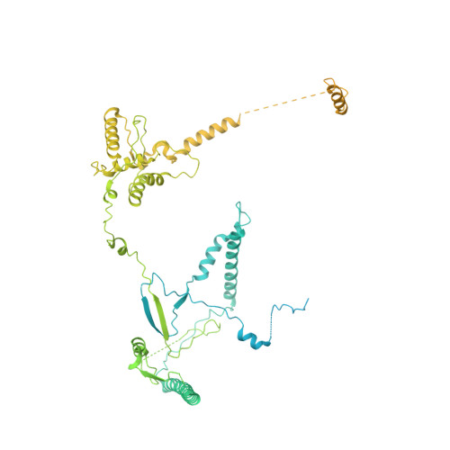
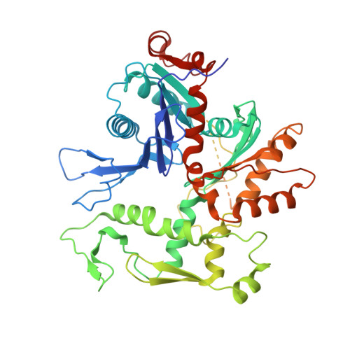
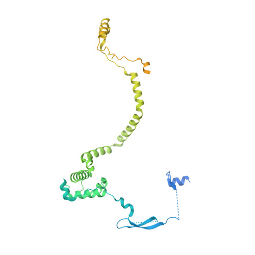
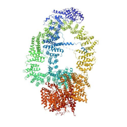
Structure of the NuA4 acetyltransferase complex bound to the nucleosome.

Qu, K.Chen, K.Wang, H.Li, X.Chen, Z.

(2022) Nature 610: 569-574

  • DOI: 10.1038/s41586-022-05303-x
  • Primary Citation of Related Structures:  
    7VVU, 7VVY, 7VVZ

  • PubMed Abstract: 
  • Deoxyribonucleic acid in eukaryotes wraps around the histone octamer to form nucleosomes 1 , the fundamental unit of chromatin. The N termini of histone H4 interact with nearby nucleosomes and play an important role in the formation of high-order chromatin structure and heterochromatin silencing 2-4 ...

    Deoxyribonucleic acid in eukaryotes wraps around the histone octamer to form nucleosomes 1 , the fundamental unit of chromatin. The N termini of histone H4 interact with nearby nucleosomes and play an important role in the formation of high-order chromatin structure and heterochromatin silencing 2-4 . NuA4 in yeast and its homologue Tip60 complex in mammalian cells are the key enzymes that catalyse H4 acetylation, which in turn regulates chromatin packaging and function in transcription activation and DNA repair 5-10 . Here we report the cryo-electron microscopy structure of NuA4 from Saccharomyces cerevisiae bound to the nucleosome. NuA4 comprises two major modules: the catalytic histone acetyltransferase (HAT) module and the transcription activator-binding (TRA) module. The nucleosome is mainly bound by the HAT module and is positioned close to a polybasic surface of the TRA module, which is important for the optimal activity of NuA4. The nucleosomal linker DNA carrying the upstream activation sequence is oriented towards the conserved, transcription activator-binding surface of the Tra1 subunit, which suggests a potential mechanism of NuA4 to act as a transcription co-activator. The HAT module recognizes the disk face of the nucleosome through the H2A-H2B acidic patch and nucleosomal DNA, projecting the catalytic pocket of Esa1 to the N-terminal tail of H4 and supporting its function in selective acetylation of H4. Together, our findings illustrate how NuA4 is assembled and provide mechanistic insights into nucleosome recognition and transcription co-activation by a HAT.


    Organizational Affiliation

    Tsinghua-Peking Joint Center for Life Sciences, Beijing Advanced Innovation Center for Structural Biology, Beijing, P. R. China. zhucheng_chen@tsinghua.edu.cn.


Macromolecules
Find similar proteins by:  (by identity cutoff)  |  3D Structure
Entity ID: 1
MoleculeChainsSequence LengthOrganismDetailsImage
Chromatin modification-related protein EAF1A [auth E]1,168Saccharomyces cerevisiae S288CMutation(s): 0 
Gene Names: EAF1VID21YDR359C
UniProt
Find proteins for Q06337 (Saccharomyces cerevisiae (strain ATCC 204508 / S288c))
Explore Q06337 
Go to UniProtKB:  Q06337
Entity Groups  
Sequence Clusters30% Identity50% Identity70% Identity90% Identity95% Identity100% Identity
UniProt GroupQ06337
Protein Feature View
Expand
  • Reference Sequence
Find similar proteins by:  (by identity cutoff)  |  3D Structure
Entity ID: 2
MoleculeChainsSequence LengthOrganismDetailsImage
Actin-related protein 4B [auth F]489Saccharomyces cerevisiae S288CMutation(s): 0 
Gene Names: ARP4ACT3YJL081CJ1012
UniProt
Find proteins for P80428 (Saccharomyces cerevisiae (strain ATCC 204508 / S288c))
Explore P80428 
Go to UniProtKB:  P80428
Entity Groups  
Sequence Clusters30% Identity50% Identity70% Identity90% Identity95% Identity100% Identity
UniProt GroupP80428
Protein Feature View
Expand
  • Reference Sequence
Find similar proteins by:  (by identity cutoff)  |  3D Structure
Entity ID: 3
MoleculeChainsSequence LengthOrganismDetailsImage
ActinC [auth G]375Saccharomyces cerevisiae S288CMutation(s): 0 
Gene Names: ACT1ABY1END7YFL039C
UniProt
Find proteins for P60010 (Saccharomyces cerevisiae (strain ATCC 204508 / S288c))
Explore P60010 
Go to UniProtKB:  P60010
Entity Groups  
Sequence Clusters30% Identity50% Identity70% Identity90% Identity95% Identity100% Identity
UniProt GroupP60010
Protein Feature View
Expand
  • Reference Sequence
Find similar proteins by:  (by identity cutoff)  |  3D Structure
Entity ID: 4
MoleculeChainsSequence LengthOrganismDetailsImage
SWR1-complex protein 4D [auth K]476Saccharomyces cerevisiae S288CMutation(s): 0 
Gene Names: SWC4EAF2GOD1YGR002C
UniProt
Find proteins for P53201 (Saccharomyces cerevisiae (strain ATCC 204508 / S288c))
Explore P53201 
Go to UniProtKB:  P53201
Entity Groups  
Sequence Clusters30% Identity50% Identity70% Identity90% Identity95% Identity100% Identity
UniProt GroupP53201
Protein Feature View
Expand
  • Reference Sequence
Find similar proteins by:  (by identity cutoff)  |  3D Structure
Entity ID: 5
MoleculeChainsSequence LengthOrganismDetailsImage
Enhancer of polycomb-like protein 1E [auth H]832Saccharomyces cerevisiae S288CMutation(s): 0 
Gene Names: EPL1YFL024C
UniProt
Find proteins for P43572 (Saccharomyces cerevisiae (strain ATCC 204508 / S288c))
Explore P43572 
Go to UniProtKB:  P43572
Entity Groups  
Sequence Clusters30% Identity50% Identity70% Identity90% Identity95% Identity100% Identity
UniProt GroupP43572
Protein Feature View
Expand
  • Reference Sequence
Find similar proteins by:  (by identity cutoff)  |  3D Structure
Entity ID: 6
MoleculeChainsSequence LengthOrganismDetailsImage
Transcription-associated protein 1F [auth L]3,744Saccharomyces cerevisiaeMutation(s): 0 
UniProt
Find proteins for P38811 (Saccharomyces cerevisiae (strain ATCC 204508 / S288c))
Explore P38811 
Go to UniProtKB:  P38811
Entity Groups  
Sequence Clusters30% Identity50% Identity70% Identity90% Identity95% Identity100% Identity
UniProt GroupP38811
Protein Feature View
Expand
  • Reference Sequence
Small Molecules
Ligands 2 Unique
IDChainsName / Formula / InChI Key2D Diagram3D Interactions
ATP (Subject of Investigation/LOI)
Query on ATP

Download Ideal Coordinates CCD File 
H [auth F],
J [auth G]
ADENOSINE-5'-TRIPHOSPHATE
C10 H16 N5 O13 P3
ZKHQWZAMYRWXGA-KQYNXXCUSA-N
 Ligand Interaction
MG (Subject of Investigation/LOI)
Query on MG

Download Ideal Coordinates CCD File 
G [auth F],
I [auth G]
MAGNESIUM ION
Mg
JLVVSXFLKOJNIY-UHFFFAOYSA-N
 Ligand Interaction
Experimental Data & Validation

Experimental Data

  • Method: ELECTRON MICROSCOPY
  • Resolution: 3.10 Å
  • Aggregation State: PARTICLE 
  • Reconstruction Method: SINGLE PARTICLE 

Structure Validation

View Full Validation Report


Entry History & Funding Information

Deposition Data


Funding OrganizationLocationGrant Number
Not funded--

Revision History  (Full details and data files)

  • Version 1.0: 2022-08-10
    Type: Initial release
  • Version 1.1: 2022-10-26
    Changes: Database references
  • Version 1.2: 2022-11-02
    Changes: Database references