7ZK2 | pdb_00007zk2

Crystal Structure of human Brachyury G177D variant in complex with CSC027898502


Experimental Data Snapshot

  • Method: X-RAY DIFFRACTION
  • Resolution: 1.60 Å
  • R-Value Free: 
    0.233 (Depositor), 0.230 (DCC) 
  • R-Value Work: 
    0.206 (Depositor), 0.210 (DCC) 
  • R-Value Observed: 
    0.207 (Depositor) 

wwPDB Validation   3D Report Full Report


Ligand Structure Quality Assessment 

Created with Raphaël 2.3.0Worse 01 BetterLigand structure goodness of fit to experimental dataBest fitted IU4Click on this verticalbar to view details

This is version 2.1 of the entry. See complete history


Literature

Structural insights into human brachyury DNA recognition and discovery of progressible binders for cancer therapy.

Newman, J.A.Gavard, A.E.Imprachim, N.Aitkenhead, H.Sheppard, H.E.Te Poele, R.Clarke, P.A.Hossain, M.A.Temme, L.Oh, H.J.Wells, C.I.Davis-Gilbert, Z.W.Workman, P.Gileadi, O.Drewry, D.H.

(2025) Nat Commun 16: 1596-1596

  • DOI: https://doi.org/10.1038/s41467-025-56213-1
  • Primary Citation of Related Structures:  
    6F58, 6F59, 7ZK2, 7ZKF, 7ZL2, 8A10, 8A7N, 8CDN

  • PubMed Abstract: 

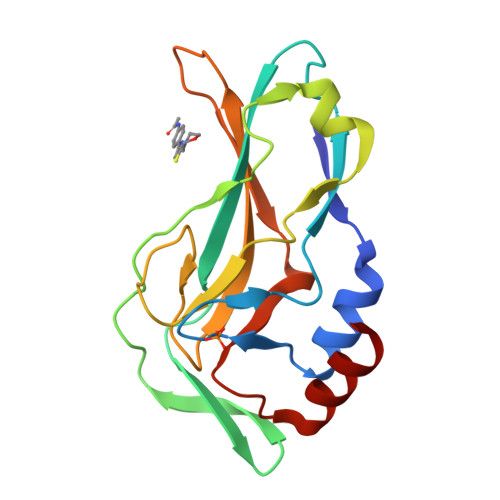
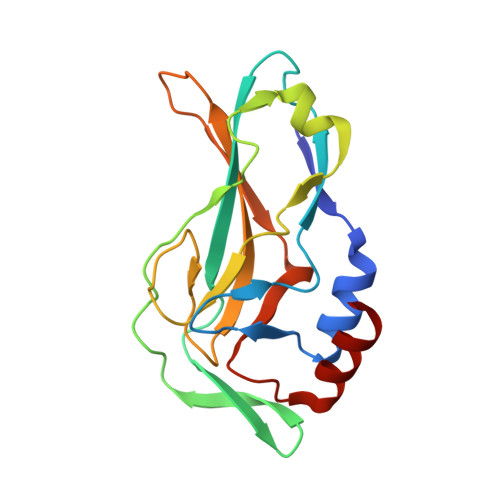
    Brachyury is a transcription factor that plays an essential role in tumour growth of the rare bone cancer chordoma and is implicated in other solid tumours. Brachyury is minimally expressed in healthy tissues, making it a potential therapeutic target. Unfortunately, as a ligandless transcription factor, brachyury has historically been considered undruggable. To investigate direct targeting of brachyury by small molecules, we determine the structure of human brachyury both alone and in complex with DNA. The structures provide insights into DNA binding and the context of the chordoma associated G177D variant. We use crystallographic fragment screening to identify hotspots on numerous pockets on the brachyury surface. Finally, we perform follow-up chemistry on fragment hits and describe the progression of a thiazole chemical series into binders with low µM potency. Thus we show that brachyury is ligandable and provide an example of how crystallographic fragment screening may be used to target protein classes that are difficult to address using other approaches.


  • Organizational Affiliation

    Centre for Medicines Discovery, University of Oxford, Oxford, UK. Joseph.newman@cmd.ox.ac.uk.


Macromolecules
Find similar proteins by:  (by identity cutoff)  |  3D Structure
Entity ID: 1
MoleculeChains Sequence LengthOrganismDetailsImage
T-box transcription factor T172Homo sapiensMutation(s): 1 
Gene Names: TBXTT
UniProt & NIH Common Fund Data Resources
Find proteins for O15178 (Homo sapiens)
Explore O15178 
Go to UniProtKB:  O15178
PHAROS:  O15178
GTEx:  ENSG00000164458 
Entity Groups  
Sequence Clusters30% Identity50% Identity70% Identity90% Identity95% Identity100% Identity
UniProt GroupO15178
Sequence Annotations
Expand
  • Reference Sequence
Experimental Data & Validation

Experimental Data

  • Method: X-RAY DIFFRACTION
  • Resolution: 1.60 Å
  • R-Value Free:  0.233 (Depositor), 0.230 (DCC) 
  • R-Value Work:  0.206 (Depositor), 0.210 (DCC) 
  • R-Value Observed: 0.207 (Depositor) 
Space Group: H 3 2
Unit Cell:
Length ( Å )Angle ( ˚ )
a = 99.92α = 90
b = 99.92β = 90
c = 98.68γ = 120
Software Package:
Software NamePurpose
PHENIXrefinement
XDSdata reduction
XDSdata scaling
REFMACphasing

Structure Validation

View Full Validation Report


Ligand Structure Quality Assessment 

Created with Raphaël 2.3.0Worse 01 BetterLigand structure goodness of fit to experimental dataBest fitted IU4Click on this verticalbar to view details

Entry History & Funding Information

Deposition Data


Funding OrganizationLocationGrant Number
The Mark FoundationUnited States--

Revision History  (Full details and data files)

  • Version 1.0: 2022-06-22
    Type: Initial release
  • Version 2.0: 2024-06-19
    Changes: Data collection, Non-polymer description
  • Version 2.1: 2025-02-26
    Changes: Database references, Structure summary