8PMR | pdb_00008pmr

NADase from Aspergillus fumigatus with mutated calcium binding motif (D219A/E220A)


Experimental Data Snapshot

  • Method: X-RAY DIFFRACTION
  • Resolution: 1.94 Å
  • R-Value Free: 
    0.216 (Depositor), 0.220 (DCC) 
  • R-Value Work: 
    0.182 (Depositor), 0.180 (DCC) 
  • R-Value Observed: 
    0.184 (Depositor) 

Starting Model: experimental
View more details

wwPDB Validation   3D Report Full Report


Ligand Structure Quality Assessment 

Created with Raphaël 2.3.0Worse 01 BetterLigand structure goodness of fit to experimental dataBest fitted NAGClick on this verticalbar to view details

This is version 1.2 of the entry. See complete history


Literature

Novel Calcium-Binding Motif Stabilizes and Increases the Activity of Aspergillus fumigatus Ecto-NADase.

Ferrario, E.Kallio, J.P.Stromland, O.Ziegler, M.

(2023) Biochemistry 62: 3293-3302

  • DOI: https://doi.org/10.1021/acs.biochem.3c00360
  • Primary Citation of Related Structures:  
    8PMR, 8PMS

  • PubMed Abstract: 

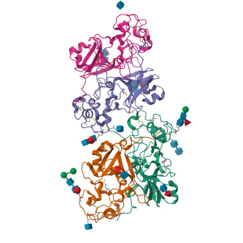
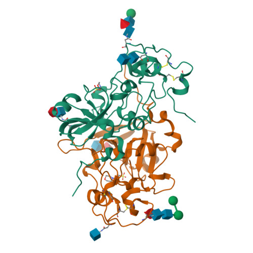
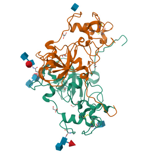
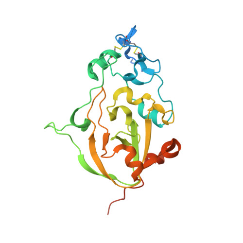
    Nicotinamide adenine dinucleotide (NAD) is an essential molecule in all kingdoms of life, mediating energy metabolism and cellular signaling. Recently, a new class of highly active fungal surface NADases was discovered. The enzyme from the opportunistic human pathogen Aspergillus fumigatus was thoroughly characterized. It harbors a catalytic domain that resembles that of the tuberculosis necrotizing toxin from Mycobacterium tuberculosis , which efficiently cleaves NAD + to nicotinamide and ADP-ribose, thereby depleting the dinucleotide pool. Of note, the A. fumigatus NADase has an additional Ca 2+ -binding motif at the C-terminus of the protein. Despite the presence of NADases in several fungal divisions, the Ca 2+ -binding motif is uniquely found in the Eurotiales order, which contains species that have immense health and economic impacts on humans. To identify the potential roles of the metal ion-binding site in catalysis or protein stability, we generated and characterized A. fumigatus NADase variants lacking the ability to bind calcium. X-ray crystallographic analyses revealed that the mutation causes a drastic and dynamic structural rearrangement of the homodimer, resulting in decreased thermal stability. Even though the calcium-binding site is at a long distance from the catalytic center, the structural reorganization upon the loss of calcium binding allosterically alters the active site, thereby negatively affecting NAD-glycohydrolase activity. Together, these findings reveal that this unique calcium-binding site affects the protein fold, stabilizing the dimeric structure, but also mediates long-range effects resulting in an increased catalytic rate.


  • Organizational Affiliation

    Department of Biomedicine, University of Bergen, Jonas Lies vei 91, Bergen 5009, Norway.


Macromolecules
Find similar proteins by:  (by identity cutoff)  |  3D Structure
Entity ID: 1
MoleculeChains Sequence LengthOrganismDetailsImage
Conidial surface nicotinamide adenine dinucleotide glycohydrolase nadA
A, B, C, D
248Aspergillus fumigatus Af293Mutation(s): 2 
Gene Names: nadAAFUA_6G14470
EC: 3.2.2.5 (PDB Primary Data), 3.2.2 (PDB Primary Data)
UniProt
Find proteins for Q4WL81 (Aspergillus fumigatus (strain ATCC MYA-4609 / CBS 101355 / FGSC A1100 / Af293))
Explore Q4WL81 
Go to UniProtKB:  Q4WL81
Entity Groups  
Sequence Clusters30% Identity50% Identity70% Identity90% Identity95% Identity100% Identity
UniProt GroupQ4WL81
Glycosylation
Glycosylation Sites: 3
Sequence Annotations
Expand
  • Reference Sequence
Oligosaccharides

Help

Entity ID: 2
MoleculeChains Length2D Diagram Glycosylation3D Interactions
2-acetamido-2-deoxy-beta-D-glucopyranose-(1-4)-2-acetamido-2-deoxy-beta-D-glucopyranose
E
2N-Glycosylation
Glycosylation Resources
GlyTouCan:  G42666HT
GlyCosmos:  G42666HT
GlyGen:  G42666HT
Entity ID: 3
MoleculeChains Length2D Diagram Glycosylation3D Interactions
beta-D-mannopyranose-(1-4)-2-acetamido-2-deoxy-beta-D-glucopyranose-(1-4)-[alpha-L-fucopyranose-(1-6)]2-acetamido-2-deoxy-beta-D-glucopyranose
F
4N-Glycosylation
Glycosylation Resources
GlyTouCan:  G32152BH
GlyCosmos:  G32152BH
GlyGen:  G32152BH
Entity ID: 4
MoleculeChains Length2D Diagram Glycosylation3D Interactions
alpha-L-fucopyranose-(1-6)-2-acetamido-2-deoxy-beta-D-glucopyranose
G, I
2N-Glycosylation
Glycosylation Resources
GlyTouCan:  G86851RC
GlyCosmos:  G86851RC
GlyGen:  G86851RC
Entity ID: 5
MoleculeChains Length2D Diagram Glycosylation3D Interactions
alpha-D-mannopyranose-(1-6)-beta-D-mannopyranose-(1-4)-2-acetamido-2-deoxy-beta-D-glucopyranose-(1-4)-[alpha-L-fucopyranose-(1-6)]2-acetamido-2-deoxy-beta-D-glucopyranose
H
5N-Glycosylation
Glycosylation Resources
GlyTouCan:  G00395TQ
GlyCosmos:  G00395TQ
GlyGen:  G00395TQ
Entity ID: 6
MoleculeChains Length2D Diagram Glycosylation3D Interactions
2-acetamido-2-deoxy-beta-D-glucopyranose-(1-4)-[alpha-L-fucopyranose-(1-6)]2-acetamido-2-deoxy-beta-D-glucopyranose
J, K, L
3N-Glycosylation
Glycosylation Resources
GlyTouCan:  G21290RB
GlyCosmos:  G21290RB
GlyGen:  G21290RB
Small Molecules
Ligands 5 Unique
IDChains Name / Formula / InChI Key2D Diagram3D Interactions
NAG
Query on NAG

Download Ideal Coordinates CCD File 
N [auth B],
P [auth C],
T [auth D]
2-acetamido-2-deoxy-beta-D-glucopyranose
C8 H15 N O6
OVRNDRQMDRJTHS-FMDGEEDCSA-N
MES
Query on MES

Download Ideal Coordinates CCD File 
M [auth A],
O [auth B]
2-(N-MORPHOLINO)-ETHANESULFONIC ACID
C6 H13 N O4 S
SXGZJKUKBWWHRA-UHFFFAOYSA-N
PEG
Query on PEG

Download Ideal Coordinates CCD File 
S [auth C]DI(HYDROXYETHYL)ETHER
C4 H10 O3
MTHSVFCYNBDYFN-UHFFFAOYSA-N
GOL
Query on GOL

Download Ideal Coordinates CCD File 
Q [auth C],
R [auth C],
U [auth D],
W [auth D]
GLYCEROL
C3 H8 O3
PEDCQBHIVMGVHV-UHFFFAOYSA-N
EDO
Query on EDO

Download Ideal Coordinates CCD File 
V [auth D]1,2-ETHANEDIOL
C2 H6 O2
LYCAIKOWRPUZTN-UHFFFAOYSA-N
Experimental Data & Validation

Experimental Data

  • Method: X-RAY DIFFRACTION
  • Resolution: 1.94 Å
  • R-Value Free:  0.216 (Depositor), 0.220 (DCC) 
  • R-Value Work:  0.182 (Depositor), 0.180 (DCC) 
  • R-Value Observed: 0.184 (Depositor) 
Space Group: P 65
Unit Cell:
Length ( Å )Angle ( ˚ )
a = 65.897α = 90
b = 65.897β = 90
c = 488.199γ = 120
Software Package:
Software NamePurpose
PHENIXrefinement
XDSdata reduction
Aimlessdata scaling
PHASERphasing

Structure Validation

View Full Validation Report


Ligand Structure Quality Assessment 

Created with Raphaël 2.3.0Worse 01 BetterLigand structure goodness of fit to experimental dataBest fitted NAGClick on this verticalbar to view details

Entry History & Funding Information

Deposition Data


Funding OrganizationLocationGrant Number
Norwegian Research CouncilNorway302314

Revision History  (Full details and data files)

  • Version 1.0: 2023-11-15
    Type: Initial release
  • Version 1.1: 2023-12-06
    Changes: Database references
  • Version 1.2: 2024-11-06
    Changes: Structure summary