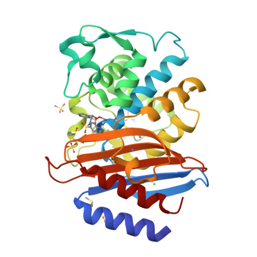
Structure of CTX-M-15 complexed with benzoxaborole AK-408
Tooke, C.L., Hinchliffe, P., Spencer, J.To be published.
Experimental Data Snapshot
Starting Model: experimental
View more details
Entity ID: 1 | |||||
|---|---|---|---|---|---|
| Molecule | Chains | Sequence Length | Organism | Details | Image |
| Beta-lactamase | 265 | Klebsiella pneumoniae | Mutation(s): 0 Gene Names: blaCTX-M-15 EC: 3.5.2.6 | 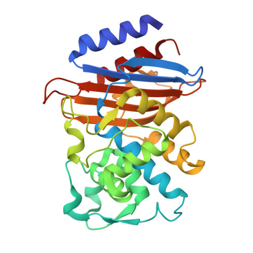 | |
UniProt | |||||
Find proteins for Q2PUH3 (Klebsiella pneumoniae) Explore Q2PUH3 Go to UniProtKB: Q2PUH3 | |||||
Entity Groups | |||||
| Sequence Clusters | 30% Identity50% Identity70% Identity90% Identity95% Identity100% Identity | ||||
| UniProt Group | Q2PUH3 | ||||
Sequence AnnotationsExpand | |||||
| |||||
| Ligands 5 Unique | |||||
|---|---|---|---|---|---|
| ID | Chains | Name / Formula / InChI Key | 2D Diagram | 3D Interactions | |
| XR3 (Subject of Investigation/LOI) Query on XR3 | B [auth A] | N-[[(3S)-1-oxidanyl-3H-2,1-benzoxaborol-3-yl]methyl]-4-[oxidanyl(oxidanylidene)-$l^{4}-azanyl]benzenesulfonamide C14 H13 B N2 O6 S GBEOHIBOUUYNIJ-HUUCEWRRSA-N |  | ||
| SO4 Query on SO4 | C [auth A], D [auth A], E [auth A], F [auth A] | SULFATE ION O4 S QAOWNCQODCNURD-UHFFFAOYSA-L |  | ||
| DMS Query on DMS | L [auth A], M [auth A] | DIMETHYL SULFOXIDE C2 H6 O S IAZDPXIOMUYVGZ-UHFFFAOYSA-N |  | ||
| CL Query on CL | G [auth A], H [auth A] | CHLORIDE ION Cl VEXZGXHMUGYJMC-UHFFFAOYSA-M |  | ||
| NA Query on NA | I [auth A], J [auth A], K [auth A] | SODIUM ION Na FKNQFGJONOIPTF-UHFFFAOYSA-N |  | ||
| Length ( Å ) | Angle ( ˚ ) |
|---|---|
| a = 44.252 | α = 90 |
| b = 45.506 | β = 90 |
| c = 117.416 | γ = 90 |
| Software Name | Purpose |
|---|---|
| PHENIX | refinement |
| DIALS | data reduction |
| Aimless | data scaling |
| PHENIX | phasing |
| Funding Organization | Location | Grant Number |
|---|---|---|
| Medical Research Council (MRC, United Kingdom) | United Kingdom | MR/T016035/1 |
RCSB PDB is hosted by

RCSB PDB is a member of the