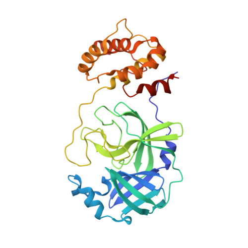
9FX7

Crystal structure of Cryo2RT SARS-CoV-2 main protease at 294K


Experimental Data Snapshot

  • Method: X-RAY DIFFRACTION
  • Resolution: 2.28 Å
  • R-Value Free: 0.244 
  • R-Value Work: 0.203 
  • R-Value Observed: 0.205 

Starting Model: experimental
View more details

wwPDB Validation   3D Report Full Report


This is version 1.2 of the entry. See complete history


Literature

Cryo2RT: a high-throughput method for room-temperature macromolecular crystallography from cryo-cooled crystals.

Huang, C.Y.Aumonier, S.Olieric, V.Wang, M.

(2024) Acta Crystallogr D Struct Biol 80: 620-628

  • DOI: https://doi.org/10.1107/S2059798324006697
  • Primary Citation of Related Structures:  
    7H56, 7H57, 7H58, 7H59, 7H5A, 7H5B, 7H5C, 7H5D, 7H5E, 7H5F, 7H5G, 7H5H, 7H5I, 7H5J, 7H5K, 7H5L, 7H5M, 7H5N, 7H5O, 7H5P, 7H5Q, 7H5R, 7H5S, 7H5T, 7H5U, 7H5V, 7H5W, 7H5X, 7H5Y, 7H5Z, 9FX4, 9FX5, 9FX6, 9FX7

  • PubMed Abstract: 

    Advances in structural biology have relied heavily on synchrotron cryo-crystallography and cryogenic electron microscopy to elucidate biological processes and for drug discovery. However, disparities between cryogenic and room-temperature (RT) crystal structures pose challenges. Here, Cryo2RT, a high-throughput RT data-collection method from cryo-cooled crystals that leverages the cryo-crystallography workflow, is introduced. Tested on endothiapepsin crystals with four soaked fragments, thaumatin and SARS-CoV-2 3CL pro , Cryo2RT reveals unique ligand-binding poses, offers a comparable throughput to cryo-crystallography and eases the exploration of structural dynamics at various temperatures.


  • Organizational Affiliation

    Swiss Light Source, Center for Photon Science, Paul Scherrer Institute, Forschungsstrasse 111, 5232 Villigen PSI, Switzerland.


Macromolecules
Find similar proteins by:  (by identity cutoff)  |  3D Structure
Entity ID: 1
MoleculeChains Sequence LengthOrganismDetailsImage
3C-like proteinase nsp5
A, B
306Severe acute respiratory syndrome coronavirus 2Mutation(s): 0 
Gene Names: rep1a-1b
EC: 3.4.22.69
UniProt
Find proteins for P0DTD1 (Severe acute respiratory syndrome coronavirus 2)
Explore P0DTD1 
Go to UniProtKB:  P0DTD1
Entity Groups  
Sequence Clusters30% Identity50% Identity70% Identity90% Identity95% Identity100% Identity
UniProt GroupP0DTD1
Sequence Annotations
Expand
  • Reference Sequence
Small Molecules
Ligands 3 Unique
IDChains Name / Formula / InChI Key2D Diagram3D Interactions
SO4
Query on SO4

Download Ideal Coordinates CCD File 
E [auth A],
J [auth B]
SULFATE ION
O4 S
QAOWNCQODCNURD-UHFFFAOYSA-L
DMS
Query on DMS

Download Ideal Coordinates CCD File 
C [auth A],
D [auth A],
G [auth A],
H [auth B],
I [auth B]
DIMETHYL SULFOXIDE
C2 H6 O S
IAZDPXIOMUYVGZ-UHFFFAOYSA-N
MG
Query on MG

Download Ideal Coordinates CCD File 
F [auth A],
K [auth B]
MAGNESIUM ION
Mg
JLVVSXFLKOJNIY-UHFFFAOYSA-N
Experimental Data & Validation

Experimental Data

  • Method: X-RAY DIFFRACTION
  • Resolution: 2.28 Å
  • R-Value Free: 0.244 
  • R-Value Work: 0.203 
  • R-Value Observed: 0.205 
  • Space Group: P 21 21 21
Unit Cell:
Length ( Å )Angle ( ˚ )
a = 68.64α = 90
b = 101.558β = 90
c = 105.255γ = 90
Software Package:
Software NamePurpose
BUSTERrefinement
XDSdata reduction
XSCALEdata scaling
PHASERphasing

Structure Validation

View Full Validation Report


Entry History & Funding Information

Deposition Data


Funding OrganizationLocationGrant Number
Swiss National Science FoundationSwitzerland182369
H2020 Marie Curie Actions of the European CommissionEuropean Union884104 PSI-FELLOW-III-3i

Revision History  (Full details and data files)

  • Version 1.0: 2024-07-31
    Type: Initial release
  • Version 1.1: 2024-08-07
    Changes: Database references
  • Version 1.2: 2024-08-14
    Changes: Database references