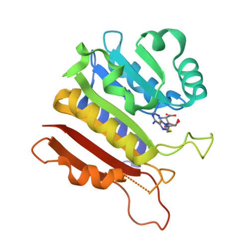
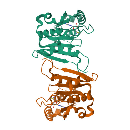
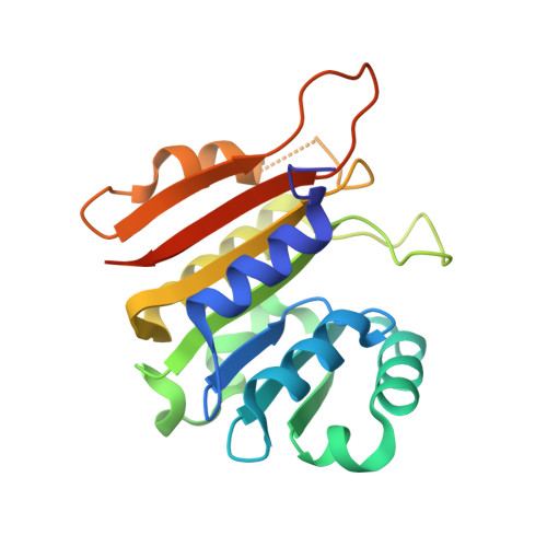
8H0T

Crystal structure of MnmM from B. subtilis complexed with SAH (1.17 A)


Experimental Data Snapshot

  • Method: X-RAY DIFFRACTION
  • Resolution: 1.17 Å
  • R-Value Free: 0.167 
  • R-Value Work: 0.135 
  • R-Value Observed: 0.136 

Starting Model: experimental
View more details

wwPDB Validation   3D Report Full Report


Ligand Structure Quality Assessment 


This is version 1.2 of the entry. See complete history


Literature

Identification of a novel 5-aminomethyl-2-thiouridine methyltransferase in tRNA modification.

Cho, G.Lee, J.Kim, J.

(2023) Nucleic Acids Res 51: 1971-1983

  • DOI: https://doi.org/10.1093/nar/gkad048
  • Primary Citation of Related Structures:  
    8H0S, 8H0T, 8H1A, 8H1B, 8H27

  • PubMed Abstract: 

    The uridine at the 34th position of tRNA, which is able to base pair with the 3'-end codon on mRNA, is usually modified to influence many aspects of decoding properties during translation. Derivatives of 5-methyluridine (xm5U), which include methylaminomethyl (mnm-) or carboxymethylaminomethyl (cmnm-) groups at C5 of uracil base, are widely conserved at the 34th position of many prokaryotic tRNAs. In Gram-negative bacteria such as Escherichia coli, a bifunctional MnmC is involved in the last two reactions of the biosynthesis of mnm5(s2)U, in which the enzyme first converts cmnm5(s2)U to 5-aminomethyl-(2-thio)uridine (nm5(s2)U) and subsequently installs the methyl group to complete the formation of mnm5(s2)U. Although mnm5s2U has been identified in tRNAs of Gram-positive bacteria and plants as well, their genomes do not contain an mnmC ortholog and the gene(s) responsible for this modification is unknown. We discovered that MnmM, previously known as YtqB, is the methyltransferase that converts nm5s2U to mnm5s2U in Bacillus subtilis through comparative genomics, gene complementation experiments, and in vitro assays. Furthermore, we determined X-ray crystal structures of MnmM complexed with anticodon stem loop of tRNAGln. The structures provide the molecular basis underlying the importance of U33-nm5s2U34-U35 as the key determinant for the specificity of MnmM.


  • Organizational Affiliation

    Department of Chemistry, Gwangju Institute of Science and Technology, Gwangju 61005, Korea.


Macromolecules
Find similar proteins by:  (by identity cutoff)  |  3D Structure
Entity ID: 1
MoleculeChains Sequence LengthOrganismDetailsImage
Putative rRNA methylase YtqB202Bacillus subtilis subsp. subtilis str. 168Mutation(s): 0 
Gene Names: ytqBBSU30490
EC: 2.1.1 (PDB Primary Data), 2.1.1.61 (UniProt)
UniProt
Find proteins for O34614 (Bacillus subtilis (strain 168))
Explore O34614 
Go to UniProtKB:  O34614
Entity Groups  
Sequence Clusters30% Identity50% Identity70% Identity90% Identity95% Identity100% Identity
UniProt GroupO34614
Sequence Annotations
Expand
  • Reference Sequence
Small Molecules
Ligands 1 Unique
IDChains Name / Formula / InChI Key2D Diagram3D Interactions
SAH (Subject of Investigation/LOI)
Query on SAH

Download Ideal Coordinates CCD File 
B [auth A]S-ADENOSYL-L-HOMOCYSTEINE
C14 H20 N6 O5 S
ZJUKTBDSGOFHSH-WFMPWKQPSA-N
Experimental Data & Validation

Experimental Data

  • Method: X-RAY DIFFRACTION
  • Resolution: 1.17 Å
  • R-Value Free: 0.167 
  • R-Value Work: 0.135 
  • R-Value Observed: 0.136 
  • Space Group: C 1 2 1
Unit Cell:
Length ( Å )Angle ( ˚ )
a = 52.555α = 90
b = 55.387β = 103.16
c = 60.592γ = 90
Software Package:
Software NamePurpose
REFMACrefinement
XDSdata reduction
STARANISOdata scaling
MOLREPphasing
MxDCdata collection
PDB_EXTRACTdata extraction
Cootmodel building

Structure Validation

View Full Validation Report


Ligand Structure Quality Assessment 


Entry History & Funding Information

Deposition Data


Funding OrganizationLocationGrant Number
National Research Foundation (NRF, Korea)Korea, Republic Of--

Revision History  (Full details and data files)

  • Version 1.0: 2023-01-25
    Type: Initial release
  • Version 1.1: 2023-03-22
    Changes: Database references
  • Version 1.2: 2023-10-25
    Changes: Data collection, Refinement description